
Kategori: Therapeutic Strategies

Spatial Pharmacology: Organelle Targeting and Condensate-Mediated Drug Discovery

Spatial Pharmacology: Organelle Targeting and Condensate-Mediated Drug Discovery

Get In Touch

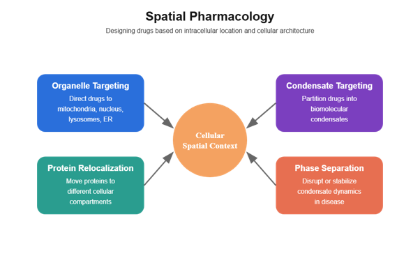
Figure: Conceptual overview of spatial pharmacology. Spatial pharmacology designs therapeutics based on intracellular location and cellular architecture. Organelle-targeted therapeutics direct drugs to specific compartments such as mitochondria or lysosomes. Protein relocalization strategies move proteins to alternative cellular locations to alter function. Condensate-targeting approaches exploit biomolecular phase separation to concentrate drugs within transcriptional or signaling condensates. Phase-separation therapeutics modulate the formation or stability of these condensates to influence disease pathways.

Executive Overview: From Molecular Binding to Spatial Control

For most of modern drug discovery, success has been defined by affinity, potency, and selectivity at the molecular level. A compound binds a protein, inhibits an enzyme, blocks a receptor. But cells are not homogeneous reaction vessels. They are spatially organized systems composed of:

  • Membrane-bound organelles
  • Dynamic trafficking networks
  • Phase-separated condensates
  • Compartment-specific microenvironments

A molecule’s biological effect depends not only on what it binds — but where it goes.

Spatial pharmacology represents a paradigm shift: therapeutics are designed with intracellular geography in mind. Instead of merely targeting a protein, drugs can:

  • Relocalize proteins to different organelles
  • Partition selectively into biomolecular condensates
  • Exploit phase separation dynamics
  • Engage bacterial or mitochondrial proteostasis machinery
  • Alter the spatial context of signaling networks

This emerging field encompasses:

  • Organelle-targeted therapeutics
  • Protein relocalization strategies
  • Condensate-mediated drug discovery
  • Phase separation therapeutics
  • Spatially restricted proteolysis systems

As our understanding of cellular architecture deepens, spatial control is becoming a new axis of drug design.

The Rise of Spatial Pharmacology

Why Location Matters in Biology

Cells compartmentalize reactions to:

  • Increase efficiency
  • Prevent crosstalk
  • Protect genomic integrity
  • Regulate signaling thresholds

Key compartments include:

  • Nucleus
  • Mitochondria
  • Endoplasmic reticulum
  • Lysosomes
  • Endosomes
  • Peroxisomes

Additionally, cells contain membraneless organelles, formed via liquid–liquid phase separation (LLPS). These condensates concentrate proteins and RNA into dynamic microdomains.

Drugs that ignore this spatial complexity may:

  • Fail to reach their intended microenvironment
  • Be sequestered away from targets
  • Disrupt unintended compartments

Spatial pharmacology embraces compartmentalization as a design principle rather than a pharmacokinetic obstacle.

Organelle-Targeted Therapeutics

Organelle-targeted therapeutics aim to direct molecules — or target proteins themselves — to specific intracellular locations.

Direct Organelle Targeting

Certain chemical motifs preferentially accumulate in specific organelles. For example:

  • Lipophilic cations accumulate in mitochondria
  • Acidic compartments trap weak bases
  • Nuclear localization sequences direct nuclear import

By engineering compounds with organelle-targeting elements, researchers can:

  • Enhance local concentration
  • Improve target engagement
  • Reduce systemic toxicity

Targeted Protein Relocalization

Beyond delivering drugs to organelles, another strategy is to move proteins themselves.

Conceptual Framework

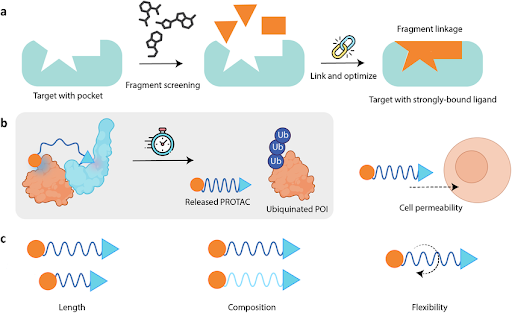
Small molecules or bifunctional chimeras can bind a protein of interest and tether it to:

  • A mitochondrial membrane
  • A nuclear export receptor
  • A lysosomal surface
  • A cytoskeletal anchor

Relocalization can:

  • Inactivate signaling pathways
  • Prevent nuclear transcriptional activity
  • Induce degradation through organelle-associated mechanisms
  • Alter metabolic flux

This represents functional knockdown without necessarily degrading the protein.

Mitochondrial Targeting as a Case Study

Mitochondria are central to:

  • Energy metabolism
  • Apoptosis regulation
  • Reactive oxygen species production

Organelle-targeted therapeutics in oncology may:

  • Induce mitochondrial stress
  • Sensitize cells to apoptosis
  • Exploit tumor metabolic rewiring

Conversely, in neurodegeneration, mitochondrial targeting can:

  • Reduce oxidative stress
  • Restore bioenergetic balance

Spatial precision improves therapeutic index.

Condensate-Mediated Drug Discovery

One of the most transformative developments in cell biology over the past decade is the discovery of biomolecular condensates.

What Are Biomolecular Condensates?

Condensates are membraneless compartments formed through liquid–liquid phase separation (LLPS). They arise when multivalent interactions among proteins and RNA cause demixing from the surrounding cytoplasm or nucleoplasm.

Examples include:

  • Nucleoli
  • Stress granules
  • P-bodies
  • Transcriptional super-enhancer condensates
  • DNA damage foci

These structures concentrate specific biomolecules while excluding others.

Why Condensates Matter for Drug Discovery

Condensates influence:

  • Gene transcription
  • RNA processing
  • Signal transduction
  • Stress responses
  • DNA repair

Drugs can:

  • Partition preferentially into condensates
  • Alter phase separation dynamics
  • Disrupt scaffold-client interactions
  • Stabilize or dissolve condensates

Thus, drug behavior is not solely defined by binding affinity — but by partitioning behavior within mesoscale cellular structures.

Drug Partitioning into Condensates

Certain small molecules concentrate within condensates due to:

  • Electrostatic interactions
  • π–π stacking
  • Intrinsically disordered region (IDR) affinity
  • RNA binding properties

Partitioning can amplify local drug concentration beyond cytoplasmic levels. However, it can also:

  • Sequester drugs away from intended targets
  • Create off-target effects within transcriptional hubs

Understanding condensate partition coefficients is becoming a critical parameter in medicinal chemistry.

Modulating Phase Separation as Therapy

Phase separation therapeutics aim to:

  • Dissolve oncogenic transcriptional condensates
  • Stabilize protective condensates
  • Prevent pathological aggregation

In oncology, super-enhancer condensates regulate oncogene expression. Disrupting their integrity can suppress transcriptional addiction.

In neurodegeneration, aberrant phase transitions may drive toxic aggregation. Modulating condensate fluidity could prevent pathological conversion.

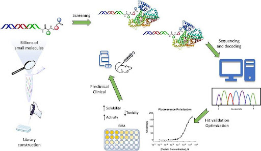
Screening Strategies in Condensate Drug Discovery

Modern screening approaches include:

  • In vitro reconstitution of phase separation
  • High-content live-cell imaging
  • Fluorescence recovery after photobleaching (FRAP)
  • Quantitative partitioning assays

Drug discovery is evolving from simple binding assays to mesoscale biophysical profiling.

Spatial Proteostasis and Bacterial Systems

Spatial pharmacology extends beyond eukaryotic organelles.

BacPROTACs: Spatial Targeting in Bacteria

BacPROTACs adapt induced proximity logic to bacterial proteostasis machinery. Rather than recruiting human E3 ligases, BacPROTACs engage bacterial proteases such as Clp systems.

Implications

  • Novel antibacterial strategies
  • Targeted degradation of resistance factors
  • Reduced reliance on enzyme inhibition

Bacterial cells lack compartmental complexity comparable to eukaryotes, but spatial organization of proteases still governs degradation efficiency.

Designing Drugs with Spatial Awareness

Spatial pharmacology requires new design principles.

Subcellular Distribution Profiling

Beyond plasma concentration, developers must assess:

  • Nuclear accumulation
  • Mitochondrial enrichment
  • Lysosomal sequestration
  • Condensate partitioning

Advanced imaging techniques now quantify intracellular distribution at high resolution.

Chemical Properties and Compartment Bias

Physicochemical parameters influence spatial behavior:

  • Charge
  • Lipophilicity
  • Polar surface area
  • Hydrogen bonding capacity

Medicinal chemists must balance permeability with compartment selectivity.

Temporal Dynamics

Spatial localization is dynamic. Stress, cell cycle stage, and signaling events alter compartment architecture.

A drug may behave differently in:

  • Hypoxic tumors
  • Neurons vs hepatocytes
  • Inflamed tissues

Time-resolved spatial profiling becomes essential.

Disease Applications of Spatial Pharmacology

Oncology

Cancer cells often reorganize spatial architecture:

  • Super-enhancer condensates drive oncogene transcription
  • DNA repair condensates maintain genomic instability
  • Metabolic enzymes relocalize under stress

Spatial pharmacology strategies may:

  • Disrupt transcriptional condensates
  • Relocalize oncogenic proteins
  • Target mitochondrial vulnerabilities

Neurodegeneration

Many neurodegenerative diseases involve:

  • Protein aggregation
  • Stress granule dysfunction
  • Aberrant phase transitions

Condensate drug discovery may prevent pathological solidification of liquid compartments.

Organelle-targeted therapeutics may protect:

  • Mitochondria
  • Lysosomes
  • Synaptic compartments

Infectious Disease

Pathogens manipulate host spatial organization:

  • Viral replication factories
  • Bacterial inclusion bodies

Spatially aware drugs could disrupt these specialized compartments.

Metabolic and Rare Diseases

Organelle dysfunction underlies:

  • Mitochondrial disorders
  • Lysosomal storage diseases
  • Peroxisomal disorders

Targeted delivery to affected organelles may improve therapeutic efficacy.

Resistance and Adaptive Remodeling

Cells adapt spatial architecture under drug pressure. Potential resistance mechanisms include:

  • Condensate remodeling
  • Organelle biogenesis changes
  • Compensatory trafficking pathways
  • Altered partitioning behavior

Understanding spatial plasticity is critical for durable therapy.

Integration with Other Modalities

Spatial pharmacology intersects with:

  • Targeted protein degradation
  • Synthetic lethality
  • RNA-targeted therapeutics
  • Immunotherapy

For example:

  • Degraders may preferentially act within condensates
  • Synthetic lethal vulnerabilities may arise from organelle stress
  • RNA-targeted drugs may accumulate in nuclear condensates

Future therapeutics may combine spatial targeting with induced proximity.

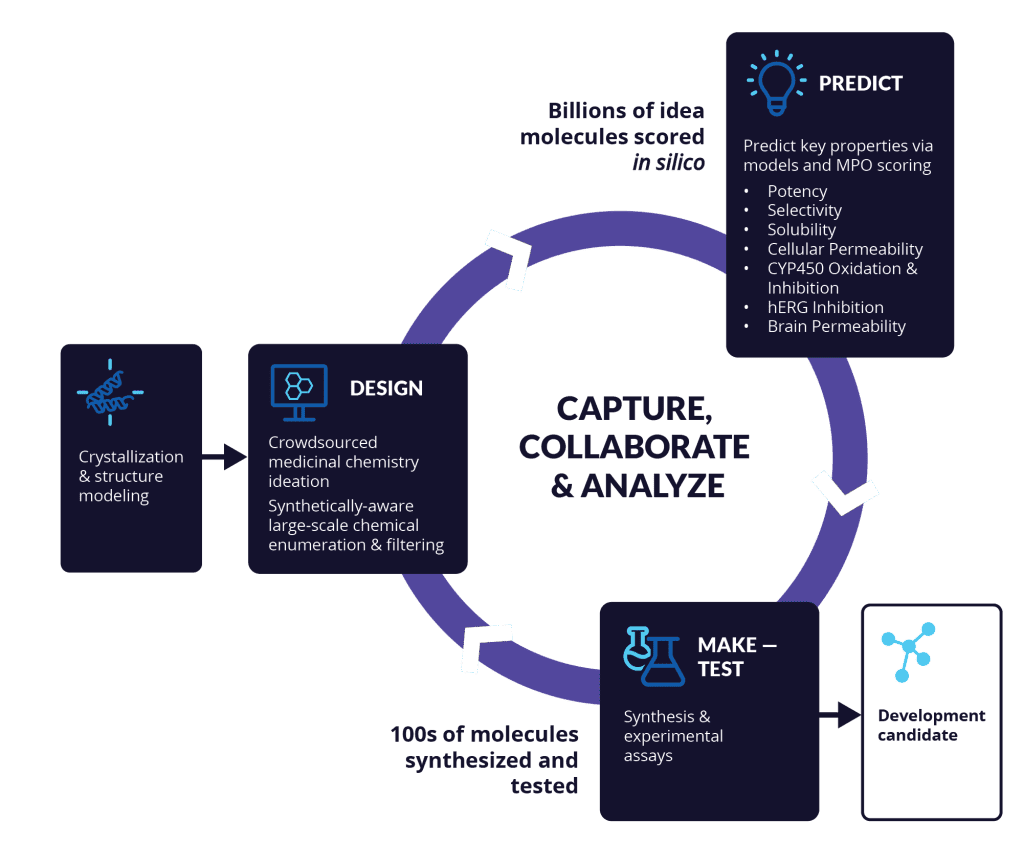
The Role of AI and Computational Modeling

Advances in machine learning enable:

  • Prediction of phase separation propensity
  • Modeling of drug partition coefficients
  • Simulation of organelle localization
  • Integration of multi-omic spatial datasets

Computational spatial pharmacology may soon guide medicinal chemistry from the earliest stages.

The Commercial Landscape

Biotech innovation in spatial pharmacology is accelerating. Emerging companies focus on:

  • Condensate-disrupting compounds
  • Organelle-targeted degraders
  • Spatially restricted proteostasis modulation
  • Mesoscale screening platforms

Pharmaceutical interest is rising because:

  • Spatial targeting expands the druggable proteome
  • Novel mechanisms create differentiation
  • Biomolecular condensates represent underexploited targets

Future Directions in Spatial Pharmacology

The next frontier may include:

  • Multi-compartment targeting chimeras
  • Programmable relocalization systems
  • Condensate-selective degraders
  • Organelle-specific E3 ligase atlases
  • Spatially resolved precision oncology

As spatial multi-omics advances, vulnerability maps may include not just gene expression — but subcellular context.

Conclusion: The Third Dimension of Drug Discovery

Traditional pharmacology operates in two dimensions:

  • Molecular affinity
  • Target selectivity

Spatial pharmacology introduces the third:

  • Intracellular location

Organelle-targeted therapeutics and condensate-mediated drug discovery redefine what it means to engage a target. The effectiveness of a drug may depend not only on binding strength, but on its ability to navigate cellular geography.

By integrating knowledge of:

  • Organelle biology
  • Phase separation
  • Intracellular trafficking
  • Mesoscale organization

drug discovery becomes not just chemistry and biology — but cellular architecture engineering.

The future of medicine may hinge on mastering this spatial dimension. In the coming decade, spatial pharmacology is poised to transform how we conceptualize therapeutic intervention — from blocking molecular function to orchestrating cellular organization itself.

Do you have an inquiry?

Contact us today and explore partnership opportunities.

Get In Touch

Related Services

Service
Small molecule drug discovery for even hard-to-drug targets – identify inhibitors, binders and modulators
Molecular Glue Direct
PPI Inhibitor Direct
Integral membrane proteins
Specificity Direct – multiplexed screening of target and anti-targets
Express – optimized for fast turn – around-time
Snap – easy, fast, and affordable

Continue reading

Next-Generation Targeted Protein Degradation: Beyond Classical PROTACs

Next-Generation Targeted Protein Degradation: Beyond Classical PROTACs

Get In Touch

Figure: Evolution of induced proximity therapeutics beyond classical PROTACs. Classical PROTACs recruit an E3 ubiquitin ligase to a target protein to induce degradation. Next-generation modalities expand this principle: RIPTACs create context-dependent proximity for tumor-selective lethality; DUBTACs stabilize proteins by recruiting deubiquitinases; PHOTACs and pcPROTACs enable light-controlled degradation; VIPER-TACs leverage viral ligases to broaden substrate scope; and PROxAb systems use antibody delivery to achieve cell-type-specific degrader activation.

Executive Overview

Targeted protein degradation (TPD) has reshaped modern drug discovery by shifting the paradigm from occupancy-driven pharmacology to event-driven pharmacology. Rather than inhibiting protein function through sustained binding, degraders trigger the removal of disease-driving proteins from the cell entirely. This catalytic mode of action enables suppression of scaffolding proteins, transcription factors, and other historically “undruggable” targets.

First-generation PROTACs (proteolysis-targeting chimeras) demonstrated that small molecules can recruit E3 ubiquitin ligases to a target protein, inducing ubiquitination and proteasomal degradation. Clinical progress in oncology validated the approach. However, the field has rapidly evolved beyond classical PROTAC architectures.

Today’s next-generation modalities expand induced proximity in multiple dimensions:

  • Context-dependent proximity (RIPTACs)
  • Targeted protein stabilization (DUBTACs)
  • Light-controlled degraders (PHOTACs, pcPROTACs)
  • Virus-inspired proximity systems (VIPER-TACs)
  • Antibody-delivered degraders (PROxAb)

Collectively, these approaches redefine TPD as a broader class of induced proximity therapeutics. The future is no longer just about degradation — it is about programmable control of protein fate, function, and location.

The Induced Proximity Paradigm

From Inhibition to Event-Driven Pharmacology

Traditional small molecules rely on occupancy: a drug must remain bound to exert effect. In contrast, degraders operate catalytically. Once a target is ubiquitinated and committed to degradation, the degrader molecule can, in principle, dissociate and engage another copy of the protein.

This creates several advantages:

  • Reduced dependence on high-affinity binding
  • Ability to eliminate scaffolding functions
  • Suppression of both enzymatic and non-enzymatic activity
  • Potential to overcome resistance mutations

At the core of TPD is induced proximity — the deliberate forced interaction between two proteins that would not normally engage. In classical PROTACs, the two components are:

  1. The target protein
  2. An E3 ubiquitin ligase

A heterobifunctional molecule brings them together, forming a ternary complex that drives ubiquitination.

However, induced proximity is not limited to E3 ligases or degradation. The same logic can recruit:

  • Deubiquitinases
  • Kinases
  • Autophagy initiators
  • RNases
  • Viral ligases
  • Conditional control elements

This conceptual shift is what defines the next wave of innovation.

RIPTACs: Context-Dependent Induced Proximity

Regulated Induced Proximity Targeting Chimeras (RIPTACs) extend the logic of proximity to create tumor-selective lethality without necessarily degrading the target.

Mechanistic Principle

RIPTACs bind:

  • A broadly expressed essential protein
  • A tumor-restricted or overexpressed protein

By forcing proximity, the essential protein becomes functionally inactivated — but only in cells expressing the tumor-specific partner. This produces synthetic-lethal-like selectivity without directly targeting mutated genes.

Why This Matters

Many essential proteins are poor therapeutic targets due to systemic toxicity. RIPTACs introduce a spatial and contextual filter: only cells co-expressing both proteins are affected.

This approach offers:

  • Precision oncology selectivity
  • Potential activity against non-mutated drivers
  • Reduced reliance on enzymatic inhibition

Challenges

  • Identifying suitable tumor-restricted anchors
  • Avoiding off-tumor expression
  • Managing adaptive rewiring

RIPTACs illustrate how induced proximity can move beyond degradation into conditional functional rewiring.

DUBTACs: Targeted Protein Stabilization

While most proximity-based drugs remove proteins, DUBTACs (Deubiquitinase-Targeting Chimeras) reverse this logic.

Mechanism

DUBTACs recruit a deubiquitinase (DUB) to a ubiquitinated target protein. This removes ubiquitin chains and rescues the protein from degradation.

Why Stabilization Matters

Many diseases arise from insufficient protein levels:

  • Tumor suppressor loss
  • Haploinsufficiency disorders
  • Misfolding-related degradation

Instead of inhibiting E3 ligases globally (which risks toxicity), DUBTACs localize deubiquitination to a specific protein.

Applications

  • Stabilizing mutant but functional tumor suppressors
  • Correcting degradation-prone variants
  • Potential rare disease interventions

Key Challenges

  • DUB selectivity and off-target deubiquitination
  • Achieving productive ternary complexes
  • Avoiding interference with endogenous ubiquitin signaling

DUBTACs demonstrate that induced proximity is not inherently degradative — it can restore function.

Conditional and Controllable Degraders

Precision control is an emerging frontier. Light-responsive degraders introduce temporal and spatial specificity.

PHOTACs: Photoswitchable PROTACs

PHOTACs incorporate photoswitchable moieties (e.g., azobenzene-like systems) that toggle between active and inactive conformations upon light exposure.

This allows:

  • Reversible activation
  • Localized degradation
  • Dynamic pathway interrogation

Potential applications include neuroscience and developmental biology, where temporal control is critical.

pcPROTACs: Photocaged PROTACs

Photocaged PROTACs remain inactive until light exposure removes a blocking group. Unlike PHOTACs, activation is typically irreversible.

Advantages

  • Clean on/off state
  • Reduced systemic activity
  • High spatiotemporal precision

PHICS and PHORCS

These systems extend light control beyond degradation into proximity-driven signaling or reversible protein complex formation.

Rather than simply removing proteins, they allow:

  • Light-triggered protein assembly
  • Signal rewiring
  • Controlled functional modulation

Collectively, these technologies move TPD toward precision pharmacology.

VIPER-TACs: Viral-Inspired Proximity Systems

Viruses encode E3 ligases and hijack host ubiquitin systems to eliminate immune regulators. VIPER-TAC strategies leverage viral ligase logic to expand the degrader toolbox.

Why Viral Ligases?

  • Distinct substrate preferences
  • Unique regulatory interfaces
  • Potential to target proteins inaccessible to canonical ligases

Applications

  • Antiviral therapies
  • Immune modulation
  • Novel substrate scope expansion

The challenge lies in specificity and safety — viral machinery must be carefully engineered to avoid unintended degradation.

PROxAb: Antibody-Delivered Degraders

One of the major limitations of small-molecule degraders is systemic exposure and tissue distribution. PROxAb platforms combine:

  • Targeting antibodies
  • Cleavable linkers
  • Intracellular degrader payloads

How It Works

  1. Antibody binds a cell-surface antigen
  2. Complex internalizes
  3. Linker cleaves intracellularly
  4. Released PROTAC engages intracellular target

This approach mirrors antibody-drug conjugates (ADCs), but instead of cytotoxic payloads, the cargo is a degrader.

Advantages

  • Cell-type specificity
  • Reduced systemic toxicity
  • Expanded therapeutic window

Technical Hurdles

  • Endosomal escape efficiency
  • Linker stability
  • Drug-to-antibody ratio optimization

PROxAb represents convergence between biologics and TPD.

Design Principles Across Next-Gen Modalities

Despite architectural diversity, common design rules apply.

Ternary Complex Cooperativity

Productive proximity requires:

  • Favorable protein-protein interactions
  • Proper geometry
  • Dynamic stability

Cooperative ternary complex formation often predicts degradation efficiency better than binary binding affinity.

Ligase or Enzyme Selection

Choice of recruited partner dictates:

  • Tissue specificity
  • Resistance pathways
  • Safety profile

Emerging strategies expand beyond CRBN and VHL to tissue-restricted ligases.

Linker Architecture

Linkers determine:

  • Flexibility vs rigidity
  • Orientation of interacting surfaces
  • Cellular permeability

Structure-guided design and AI modeling increasingly guide optimization.

Contextual Selectivity

Next-generation systems incorporate:

  • Tumor-restricted expression
  • Light activation
  • Antibody delivery
  • Conditional engagement

Selectivity is shifting from binding specificity to biological context specificity.

Clinical and Translational Landscape

Targeted protein degradation has moved from conceptual novelty to clinical reality, particularly in oncology. Next-generation modalities aim to address:

  • Solid tumor resistance
  • Central nervous system delivery
  • Rare genetic disorders
  • Immune modulation

Several biotech companies are now expanding pipelines into:

  • Tissue-restricted ligase discovery
  • Autophagy-based degradation
  • RNA-targeted proximity systems

As the field matures, differentiation increasingly depends on:

  • Ligase access
  • Conditional control
  • Safety margins
  • Novel substrate space

Resistance and Evolution of the Field

Resistance mechanisms include:

  • E3 ligase downregulation
  • Target mutation
  • Adaptive rewiring
  • Proteostasis compensation

Next-generation platforms attempt to preempt resistance through:

  • Alternative ligases
  • Conditional activation
  • Multi-target strategies

Combinatorial proximity drugs may represent a future frontier.

The Future of Induced Proximity Therapeutics

The evolution of targeted protein degradation reveals a broader principle: drug discovery is becoming programmable biology.

Future directions include:

  • Multi-functional chimeras that degrade one protein while stabilizing another
  • Tissue-restricted ligase atlases
  • AI-designed ternary complexes
  • Integration with synthetic lethality frameworks
  • Expansion beyond proteins to RNA and chromatin complexes

Ultimately, induced proximity is not a single modality — it is a design philosophy.

The next decade will likely see:

  • More precise control
  • Broader target classes
  • Increased integration with biologics
  • Spatial and temporal programmability

Targeted protein degradation began as a clever workaround for undruggable proteins. It is now evolving into a foundational technology platform capable of reshaping therapeutic intervention at a systems level.

The era beyond classical PROTACs has begun — and it is defined not by degradation alone, but by the controlled orchestration of molecular proximity.

Do you have an inquiry?

Contact us today and explore partnership opportunities.

Get In Touch

Related Services

Service
Small molecule drug discovery for even hard-to-drug targets – identify inhibitors, binders and modulators
Molecular Glue Direct
PPI Inhibitor Direct
Integral membrane proteins
Specificity Direct – multiplexed screening of target and anti-targets
Express – optimized for fast turn – around-time
Snap – easy, fast, and affordable

Continue reading

Synthetic Lethality and Context-Dependent Cancer Targeting

Synthetic Lethality and Context-Dependent Cancer Targeting

Get In Touch

Figure: Conceptual model of synthetic lethality in cancer therapy. In normal cells, redundant pathways allow survival when either gene is inhibited. In cancer cells, mutation of one gene creates dependency on a compensatory pathway. Targeting this remaining pathway produces synthetic lethality, selectively killing tumor cells while sparing normal tissue. Clinically relevant examples include WRN inhibition in MSI-high tumors and PRMT5 targeting in MTAP-deleted cancers.

Executive Overview: From Oncogene Inhibition to Dependency Targeting

For decades, oncology drug discovery focused on inhibiting activated oncogenes — mutant kinases, amplified receptors, dysregulated signaling enzymes. While transformative in many cases, this strategy has intrinsic limits:

  • Not all cancers harbor targetable oncogenic mutations
  • Many driver proteins lack druggable pockets
  • Tumors evolve resistance through pathway rewiring

Synthetic lethality offers a fundamentally different approach. Instead of directly targeting the mutation, it exploits context-specific dependencies created by that mutation.

In simple terms: two genes are synthetically lethal if loss of either alone is tolerated, but loss of both is lethal.

Cancer cells, by virtue of their mutations, often become dependent on backup pathways that normal cells do not require. Synthetic lethality drug discovery aims to identify and therapeutically exploit these vulnerabilities.

The modern era of synthetic lethality encompasses:

  • Genome-scale CRISPR screening
  • Context-dependent essentiality mapping
  • Precision biomarker-driven trials
  • Integration with induced proximity therapeutics

Key exemplars include:

  • WRN inhibitors in MSI-high cancers
  • PRMT5 targeting in MTAP-deleted tumors
  • Context-dependent induced proximity strategies

This pillar article explores how synthetic lethality is reshaping precision oncology.

Foundations of Synthetic Lethality

Historical Context

The concept of synthetic lethality originated in classical genetics. In model organisms, researchers observed that deletion of either Gene A or Gene B was tolerated, but deletion of both caused death.

In cancer biology, the idea became clinically validated with PARP inhibitors in BRCA-mutated tumors — demonstrating that therapeutic synthetic lethality could be achieved in patients.

This success shifted oncology strategy from:

  • “Target the mutation”

to:

  • “Target what the mutation makes essential.”

Feasibility study: 2-4 weeks
Screening: 4-8 weeks

Why Cancer Cells Are Vulnerable

Cancer cells accumulate:

  • DNA repair defects
  • Replication stress
  • Metabolic rewiring
  • Proteotoxic stress
  • Chromosomal instability

These alterations create dependencies on compensatory pathways. While normal cells retain redundancy, tumor cells often operate at the edge of viability.

Synthetic lethality drug discovery aims to identify these edge conditions.

Precision Oncology Vulnerabilities: Mapping Contextual Dependencies

Modern synthetic lethality relies heavily on:

  • CRISPR knockout screens
  • RNAi dependency maps
  • Multi-omic profiling
  • Patient-derived models

Large-scale datasets now catalog gene essentiality across hundreds of cancer cell lines. These maps reveal:

  • Lineage-specific vulnerabilities
  • Mutation-specific dependencies
  • Epigenetic context effects

The challenge lies in translating these genetic interactions into druggable targets with sufficient therapeutic windows.

WRN Dependency in MSI-High Tumors

One of the most compelling recent examples of synthetic lethality involves WRN helicase dependency in microsatellite instability-high (MSI-high) cancers. WRN plays a critical role in DNA replication and repair.

Biological Context: MSI-High Tumors

MSI-high cancers arise from mismatch repair (MMR) deficiency. Loss of MMR leads to:

  • Accumulation of insertion/deletion mutations
  • Replication stress
  • DNA secondary structure instability

To survive this genomic instability, MSI-high tumor cells become dependent on WRN helicase activity.

Mechanistic Basis of WRN Synthetic Lethality

WRN helps resolve:

  • DNA secondary structures
  • Replication fork stalling
  • Microsatellite-induced instability

In MMR-deficient cells, unresolved structures accumulate rapidly without WRN, leading to:

  • Chromosomal breakage
  • Mitotic catastrophe
  • Cell death

In contrast, normal MMR-proficient cells tolerate WRN inhibition more effectively.

Therapeutic Development

WRN inhibitors represent a promising strategy in MSI-high colorectal, gastric, and endometrial cancers.

Advantages include:

  • Strong genetic validation
  • Biomarker clarity (MSI-high status)
  • Potential synergy with immunotherapy

Challenges remain:

  • Achieving selective helicase inhibition
  • Avoiding toxicity in proliferative tissues
  • Managing resistance through compensatory repair pathways

WRN inhibition exemplifies synthetic lethality driven by DNA repair deficiency.

PRMT5 Dependency in MTAP-Deleted Tumors

Another well-characterized vulnerability involves PRMT5 inhibition in tumors with MTAP deletion. PRMT5 is an epigenetic regulator involved in symmetric arginine methylation. MTAP is frequently co-deleted with CDKN2A in many cancers.

Biological Context

MTAP deletion leads to accumulation of methylthioadenosine (MTA), a metabolite that partially inhibits PRMT5. This creates a state where:

  • PRMT5 activity is already compromised
  • Cancer cells operate near minimal PRMT5 function
  • Further inhibition becomes selectively lethal

Mechanistic Insight

PRMT5 regulates:

  • Splicing
  • Chromatin modification
  • Transcriptional control

MTAP-deleted cells are hypersensitive to PRMT5 inhibition because MTA accumulation lowers the functional threshold.

Therapeutic approaches include:

  • Substrate-competitive inhibitors
  • MTA-cooperative inhibitors
  • Context-selective PRMT5 targeting

Clinical Implications

MTAP deletion is common across:

  • Glioblastoma
  • Pancreatic cancer
  • Lung cancer
  • Mesothelioma

This provides a broad patient population with defined biomarker stratification.

Key development challenges:

  • Avoiding hematologic toxicity
  • Optimizing therapeutic index
  • Understanding splicing-related side effects

PRMT5–MTAP represents a metabolic-epigenetic synthetic lethal axis.

Induced Proximity and Context-Dependent Targeting

Beyond classical enzyme inhibition, induced proximity technologies are beginning to intersect with synthetic lethality strategies. RIPTACs represent one such example.

Context-Dependent Induced Proximity

RIPTACs force interaction between:

  • A broadly expressed essential protein
  • A tumor-restricted protein

The essential protein becomes functionally impaired only in cells expressing the tumor-specific anchor. This creates synthetic lethality without directly targeting a mutated gene.

Advantages

  • Expanded druggable space
  • Selectivity driven by co-expression
  • Potential targeting of non-mutant tumors

Induced proximity adds a programmable dimension to synthetic lethality.

Discovery Strategies in Synthetic Lethality Drug Discovery

CRISPR-Based Screens

Genome-wide CRISPR knockout or CRISPR interference screens identify:

  • Mutation-specific essential genes
  • Pathway dependencies
  • Synergistic vulnerabilities

High-throughput combinatorial screens accelerate mapping of lethal gene pairs.

Multi-Omic Integration

Synthetic lethality is rarely binary. Context matters:

  • Tumor lineage
  • Epigenetic state
  • Metabolic environment
  • Microenvironmental stress

Integrating genomics, transcriptomics, proteomics, and metabolomics improves predictive accuracy.

Patient Stratification

Precision oncology vulnerabilities require robust biomarkers:

  • MSI-high status for WRN
  • MTAP deletion for PRMT5
  • Specific mutation signatures

Without clear stratification, therapeutic windows collapse.

Resistance Mechanisms

As with any targeted therapy, resistance can emerge. Common resistance pathways include:

  • Restoration of lost repair functions
  • Upregulation of compensatory pathways
  • Mutation of drug target
  • Epigenetic adaptation

For example:

  • Alternative helicases may compensate for WRN inhibition
  • PRMT5 pathway rewiring may restore splicing balance

Combination strategies are often necessary to sustain efficacy.

Combination Strategies

Synthetic lethality often synergizes with:

  • Immunotherapy
  • DNA-damaging agents
  • Targeted protein degradation
  • Epigenetic modulators

For example:

  • WRN inhibition may increase tumor mutational burden and immune visibility
  • PRMT5 inhibition may sensitize tumors to splicing stress

Rational combinations are critical for durable response.

Expanding the Synthetic Lethal Landscape

Emerging directions include:

  • Targeting replication stress pathways
  • Exploiting metabolic bottlenecks
  • Synthetic lethal interactions with chromatin remodelers
  • Leveraging tumor microenvironment dependencies

AI-driven models may soon predict synthetic lethal interactions de novo.

Commercial and Translational Landscape

Synthetic lethality drug discovery has become a cornerstone of precision oncology pipelines.

Pharmaceutical interest is high because:

  • Genetic validation reduces target risk
  • Biomarker-defined populations improve trial efficiency
  • First-in-class opportunities are abundant

Companies are investing in:

  • Dependency mapping platforms
  • CRISPR-enabled discovery
  • Computational vulnerability prediction

Synthetic lethality is now integrated into mainstream oncology R&D strategy.

Future Outlook: From Mutation-Driven to Dependency-Driven Oncology

The future of cancer therapy may depend less on targeting oncogenes directly and more on targeting what cancer cells cannot live without.

Synthetic lethality reframes cancer not as a disease of activation alone, but as a disease of vulnerability.

Key future developments may include:

  • Personalized dependency maps
  • Dynamic vulnerability profiling during treatment
  • Synthetic lethal networks rather than single pairs
  • Integration with spatial and proximity-based therapeutics

Precision oncology vulnerabilities are likely to become increasingly refined as tumor heterogeneity is better understood.

Conclusion

Synthetic lethality drug discovery represents one of the most rational and genetically grounded approaches in modern oncology.

From WRN inhibitors in MSI-high cancers to PRMT5 targeting in MTAP-deleted tumors, context-specific vulnerabilities are yielding actionable therapeutic strategies.

The core insight is simple but powerful: cancer cells survive by rewiring biology — and that rewiring creates dependencies.

By systematically identifying and exploiting those dependencies, precision oncology moves beyond inhibiting drivers to dismantling survival scaffolds.

As discovery technologies mature and induced proximity tools expand the targetable space, synthetic lethality will continue to redefine how cancer is treated — not by attacking what tumors are, but by exploiting what they cannot live without.

Do you have an inquiry?

Contact us today and explore partnership opportunities.

Get In Touch

Related Services

Service
Small molecule drug discovery for even hard-to-drug targets – identify inhibitors, binders and modulators
Molecular Glue Direct
PPI Inhibitor Direct
Integral membrane proteins
Specificity Direct – multiplexed screening of target and anti-targets
Express – optimized for fast turn – around-time
Snap – easy, fast, and affordable

Continue reading

RNA-Targeted Small Molecules and Translation-Modulating Therapeutics

RNA-Targeted Small Molecules and Translation-Modulating Therapeutics

Get In Touch

Figure: Major strategies in RNA-targeted small-molecule therapeutics. RNA-targeted pharmacology acts upstream of protein synthesis. Splicing modifiers alter exon selection in pre-mRNA to reshape transcript isoforms. RIBOTACs recruit endogenous ribonucleases such as RNase L to selectively degrade disease-causing RNA transcripts. Ribosome-stalling small molecules bind within the ribosomal exit tunnel and arrest translation of specific nascent peptide sequences, suppressing protein production. Together these approaches expand drug discovery beyond proteins to the transcriptome.

Executive Overview: Expanding the Druggable Genome Upstream of Protein

For decades, small-molecule drug discovery has focused almost exclusively on proteins — particularly enzymes and receptors with well-defined binding pockets. Yet the human genome encodes far more RNA species than druggable proteins. Messenger RNAs (mRNAs), long noncoding RNAs (lncRNAs), microRNAs, repeat-expansion transcripts, and structured untranslated regions represent a vast and largely untapped regulatory landscape.

Recent advances in structural biology, RNA chemistry, and proximity-based pharmacology have catalyzed a new frontier: RNA-targeted small molecules and translation-modulating therapeutics.

Rather than inhibiting protein function after synthesis, these strategies act upstream by:

  • Modulating pre-mRNA splicing
  • Degrading disease-causing RNA selectively
  • Arresting translation of specific nascent polypeptides
  • Exploiting RNA structure as a druggable motif

The field now encompasses:

  • Splicing-modifier therapeutics
  • RIBOTACs
  • Ribosome-stalling small molecules (“interdictors”)

Collectively, these approaches redefine small-molecule pharmacology — from targeting static proteins to dynamically regulating gene expression itself.

Why RNA Is Emerging as a Druggable Target

The Limitations of Protein-Centric Drug Discovery

Many disease drivers are difficult to inhibit at the protein level:

  • Transcription factors lacking enzymatic pockets
  • Scaffolding proteins
  • Gain-of-function mutations
  • Proteins expressed at extremely high levels

Targeting RNA offers several advantages:

  • Control before translation
  • Access to noncoding regulatory molecules
  • Potential for high selectivity via structural motifs
  • Opportunity to eliminate toxic transcripts directly

Importantly, RNA structures are not random coils. Many RNAs fold into:

  • Hairpins
  • Bulges
  • Internal loops
  • G-quadruplexes
  • Pseudoknots

These structural motifs create binding surfaces analogous to protein pockets.

Splicing-Modifying Therapeutics

Pre-mRNA splicing is a tightly regulated process that removes introns and joins exons. Errors in splicing contribute to:

  • Cancer
  • Neurodegeneration
  • Genetic diseases

Small molecules can alter splice-site selection or spliceosome activity, shifting transcript isoforms in therapeutically beneficial ways.

Mechanistic Basis

Splicing modifiers typically interact with:

  • Core spliceosome components
  • RNA-protein interfaces
  • Structured RNA motifs within pre-mRNA

These interactions alter exon inclusion or exclusion.

In oncology, mutations in splicing factors (e.g., SF3B1) create vulnerabilities. Cancer cells often operate near the threshold of splicing fidelity. Small perturbations can push them into transcriptomic catastrophe.

Splicing Modifiers in Cancer

Cancer cells frequently harbor:

  • Spliceosome mutations
  • Elevated transcriptional stress
  • Increased reliance on aberrant splicing programs

Splicing modifiers can:

  • Induce exon skipping in oncogenic transcripts
  • Create neoantigens
  • Trigger apoptosis via splicing overload

This creates a therapeutic window distinct from normal cells.

Genetic Disease Applications

Beyond oncology, splicing correction has proven transformative in inherited disorders where exon misselection drives pathology.

Small molecules offer advantages over antisense oligonucleotides:

  • Oral bioavailability potential
  • Broader tissue penetration
  • Lower manufacturing complexity

However, transcriptome-wide effects remain a concern. Dose-dependent selectivity and careful biomarker development are critical.

RIBOTACs: Targeted RNA Degradation

RIBOTACs represent a catalytic approach to RNA elimination.

Conceptual Framework

RIBOTACs are bifunctional small molecules that:

  1. Bind a structured RNA target
  2. Recruit an endogenous ribonuclease (often RNase L)

This induced proximity triggers selective cleavage of the target RNA.

Unlike antisense oligonucleotides (ASOs) or siRNA, RIBOTACs:

  • Are small molecules
  • Potentially cross membranes more efficiently
  • Function catalytically
  • Exploit endogenous RNase machinery

Mechanistic Details

RNase L is normally activated in antiviral responses. RIBOTACs co-opt this system by tethering RNase L near a disease-relevant RNA.

Key design principles include:

  • High-affinity RNA binding
  • Minimal off-target RNase recruitment
  • Proper geometric alignment
  • Avoiding global RNase activation

The ternary complex — RNA + RIBOTAC + RNase — parallels PROTAC logic but in the RNA domain.

Comparison to Other RNA Therapeutics

Feature RIBOTAC ASO siRNA
Size Small molecule Oligonucleotide Oligonucleotide
Delivery complexity Moderate High High
Catalytic action Yes No Yes (RISC-mediated)
Immune activation risk Variable Moderate Moderate
Oral potential Theoretical Rare Rare
RIBOTACs may complement nucleic acid therapeutics rather than replace them.

Target Space

Potential RNA targets include:

  • Structured mRNAs
  • Oncogenic lncRNAs
  • Repeat expansion transcripts
  • Viral RNAs

Structured motifs are particularly attractive because they create binding specificity.

Ribosome-Stalling Therapeutics (“Interdictors”)

While RIBOTACs eliminate RNA, ribosome-stalling molecules act during translation.

Mechanistic Principle

Selective ribosome-stalling compounds bind within the ribosomal exit tunnel and interact with specific nascent peptide sequences. This causes:

  • Translational arrest
  • Premature termination
  • Reduced protein output

Importantly, stalling can be sequence-dependent, enabling selective targeting of specific proteins.

Distinguishing from Classical Translation Inhibitors

Traditional translation inhibitors (e.g., broad-spectrum antibiotics) globally suppress protein synthesis.

Ribosome-stalling therapeutics aim for:

  • Target-selective arrest
  • Reduced global toxicity
  • Context-dependent effects

By recognizing specific amino acid motifs emerging from the ribosome, these compounds achieve precision.

Applications in Oncology

Many oncogenic drivers are difficult to degrade post-translationally due to:

  • High expression
  • Rapid turnover
  • Structural inaccessibility

Translation arrest offers an alternative:

  • Suppress production at the source
  • Reduce protein accumulation
  • Potentially overcome degradation resistance

Challenges

  • Avoiding unintended translational stress responses
  • Managing integrated stress pathway activation
  • Ensuring sequence selectivity

High-resolution cryo-EM studies have been critical in elucidating binding mechanisms.

Design Principles in RNA-Targeted Small Molecules

RNA Structural Mapping

RNA structure is dynamic and context-dependent. Key tools include:

  • SHAPE-MaP
  • Cryo-EM
  • NMR
  • Chemical probing

Accurate structural models are essential for rational design.

Binding Selectivity

RNA is chemically similar across transcripts. Selectivity arises from:

  • Unique structural motifs
  • Bulge geometry
  • Base stacking interactions
  • Minor groove architecture

Medicinal chemistry must account for:

  • Electrostatic interactions
  • Hydrogen bonding networks
  • Conformational flexibility

Avoiding Off-Target RNA Effects

Because many RNAs share structural features, off-target risks include:

  • Unintended splicing shifts
  • Degradation of essential transcripts
  • Activation of innate immune pathways

High-throughput transcriptomic profiling is mandatory.

Induced Proximity Logic in RNA Context

Like PROTACs, RIBOTACs depend on ternary complex formation. Design parameters include:

  • Orientation constraints
  • Flexible vs rigid linkers
  • Enzyme recruitment efficiency
  • Catalytic turnover

AI-driven modeling may soon enable predictive RNA–ligand–enzyme complex design.

Therapeutic Applications

Oncology

RNA-targeted small molecules can:

  • Suppress oncogenic splice variants
  • Degrade oncogenic transcripts
  • Arrest translation of driver proteins
  • Exploit cancer-specific RNA dependencies

Tumors with splicing factor mutations are particularly vulnerable.

Repeat Expansion Disorders

Diseases caused by toxic RNA repeats (e.g., CAG, CGG expansions) are prime candidates for RIBOTAC strategies.

Selective degradation of repeat-containing transcripts could:

  • Reduce toxic RNA foci
  • Decrease aberrant protein translation
  • Modify disease course

Viral Infections

Structured viral RNAs represent attractive targets:

  • Conserved secondary structures
  • Essential replication elements
  • Reduced host homologs

RIBOTAC-like strategies could selectively degrade viral genomes.

Neurodegeneration

RNA-binding proteins and aberrant RNA splicing are central to:

  • ALS
  • Frontotemporal dementia
  • Huntington’s disease

Splicing correction and RNA degradation offer novel intervention points.

Integration with Other Modalities

RNA-targeted approaches can synergize with:

  • Targeted protein degradation
  • Synthetic lethality strategies
  • Immunotherapy
  • Gene editing

For example:

  • RNA suppression may sensitize tumors to degrader therapy
  • Splicing modulation can create neoantigens for immune targeting

The therapeutic ecosystem is increasingly combinatorial.

Clinical and Commercial Landscape

RNA-targeted drug discovery is transitioning from exploratory chemistry to translational programs.

Key enabling factors:

  • Improved RNA structural resolution
  • Better predictive modeling
  • Growing understanding of transcriptome dynamics

Investment interest reflects recognition that:

  • RNA expands the druggable universe
  • Structured RNAs provide specificity opportunities
  • Small molecules offer advantages over nucleic acids in certain contexts

Companies are building pipelines around:

  • Splicing modulation
  • RNA degraders
  • Translation regulators

Resistance Considerations

As with protein-targeted drugs, resistance may arise via:

  • RNA mutation altering structure
  • Alternative splicing compensation
  • Upregulation of parallel pathways
  • Stress response activation

Combination therapies may mitigate resistance risk.

Future Directions in RNA-Targeted Drug Discovery

The next wave of innovation may include:

  • Programmable RNA degraders
  • Multi-functional RNA-binding chimeras
  • Tissue-specific RNA targeting strategies
  • AI-guided RNA structure prediction
  • Condensate-aware RNA pharmacology

Advances in computational biology will likely accelerate identification of:

  • Druggable RNA motifs
  • Disease-specific transcript vulnerabilities
  • RNA–protein interaction hotspots

Conclusion: Rewriting the Central Dogma of Pharmacology

RNA-targeted small molecules and translation-modulating therapeutics represent a fundamental expansion of drug discovery logic.

Rather than intervening after protein synthesis, these approaches act:

  • At the transcript level
  • During translation
  • Within splicing regulation

They convert RNA from an informational intermediary into a direct pharmacological substrate.

As the field matures, RNA-targeted drug discovery may parallel — and in some cases surpass — protein-centric approaches in impact. Structured RNAs, inducible ribonuclease recruitment, and selective ribosome stalling collectively broaden the scope of what is therapeutically addressable.

The central dogma once defined biology as DNA → RNA → protein. Modern pharmacology increasingly operates upstream — at RNA — where disease signals originate.

The era of RNA-targeted small molecules has begun.

Do you have an inquiry?

Contact us today and explore partnership opportunities.

Get In Touch

Related Services

Service
Small molecule drug discovery for even hard-to-drug targets – identify inhibitors, binders and modulators
Molecular Glue Direct
PPI Inhibitor Direct
Integral membrane proteins
Specificity Direct – multiplexed screening of target and anti-targets
Express – optimized for fast turn – around-time
Snap – easy, fast, and affordable

Continue reading

Autophagy and Lysosome-Based Targeted Degradation Technologies

Autophagy and Lysosome-Based Targeted Degradation Technologies

Get In Touch

Figure: Overview of lysosome- and autophagy-based targeted degradation technologies.
Lysosomal degradation pathways expand targeted protein degradation beyond the ubiquitin–proteasome system. LYTACs and PROTABs enable clearance of extracellular and membrane proteins through receptor-mediated endocytosis and lysosomal trafficking. Intracellular autophagy-based systems include ATTECs (LC3 tethering), AUTACs (K63-linked ubiquitin autophagy signaling), AUTOTACs (p62-mediated recruitment), and ULKTACs, which initiate autophagy through ULK1 activation. Together these technologies broaden the degradable target space to include extracellular proteins, aggregates, and organelles.

Beyond the Proteasome: Why Lysosomal Pathways Matter

Targeted protein degradation (TPD) initially centered on the ubiquitin–proteasome system (UPS). Classical PROTACs demonstrated that small molecules could catalytically eliminate intracellular proteins by recruiting E3 ligases. Yet the proteasome has intrinsic constraints:

  • It primarily degrades soluble intracellular proteins
  • It struggles with large aggregates and organelles
  • It cannot directly eliminate extracellular or membrane proteins
  • Its activity can be compromised in stressed or diseased cells

These limitations have catalyzed the rise of lysosome- and autophagy-based targeted degradation technologies — modalities that harness the cell’s broader proteostasis and trafficking machinery.

Unlike the proteasome, the lysosome:

  • Degrades membrane proteins and extracellular ligands
  • Handles protein aggregates and damaged organelles
  • Integrates with endocytosis and macroautophagy
  • Provides access to previously “undruggable” spatial compartments

This expansion of degradable target space is reshaping therapeutic strategies in oncology, neurodegeneration, immunology, and rare disease.

Lysosome-Targeting Chimeras (LYTACs): Clearing the Extracellular Proteome

LYTACs represent a foundational advance in extracellular protein degradation.

Mechanistic Overview

LYTACs are bifunctional molecules that bind:

  1. An extracellular or membrane-bound target protein
  2. A lysosome-shuttling receptor (such as CI-M6PR or ASGPR)

Upon binding, the ternary complex is internalized through endocytosis and trafficked to the lysosome for degradation.

This enables:

  • Degradation of secreted cytokines
  • Removal of membrane receptors
  • Clearance of pathogenic extracellular proteins

Unlike monoclonal antibodies, which typically block function, LYTACs eliminate the target entirely.

Receptor Systems Used in LYTAC Design

CI-M6PR (Cation-Independent Mannose-6-Phosphate Receptor)

  • Broad tissue expression
  • Established lysosomal trafficking route
  • Suitable for systemic applications

ASGPR (Asialoglycoprotein Receptor)

  • Liver-restricted expression
  • Enables hepatocyte-specific degradation
  • Valuable for metabolic and liver diseases

Receptor selection determines tissue specificity and therapeutic index.

Therapeutic Applications of LYTACs

Oncology

  • Degradation of growth factor receptors
  • Removal of immune checkpoint ligands
  • Targeting shed extracellular drivers

Immunology

  • Cytokine clearance
  • Modulation of inflammatory mediators

Fibrotic Disease

  • Eliminating profibrotic secreted proteins

LYTACs extend TPD into compartments previously inaccessible to small molecules.

LYMTACs and PROTABs: Next-Generation Surface Target Clearance

Beyond classical LYTACs, newer modalities refine surface protein degradation strategies.

LYMTACs (Lysosomal Membrane Targeting Chimeras)

LYMTACs expand lysosome-directed degradation by targeting lysosomal membrane proteins or alternative internalization routes.

Key differentiators:

  • Exploit distinct lysosomal membrane trafficking pathways
  • Potentially bypass limitations of CI-M6PR dependency
  • Broaden receptor options

LYMTACs increase flexibility in receptor biology and tissue targeting.

PROTABs (Proteolysis-Targeting Antibodies)

PROTABs are antibody-based constructs designed to induce degradation of cell-surface targets.

Mechanistic logic:

  • Antibody binds membrane target
  • Engages endogenous ubiquitination or internalization machinery
  • Drives lysosomal degradation

Unlike classical antibodies that merely block receptor function, PROTABs induce complete target removal.

Advantages over mAbs:

  • Sustained target depletion
  • Reduced compensatory signaling
  • Potential to overcome resistance to receptor blockade

Selective Autophagy-Based Targeted Degradation

While LYTACs operate via endocytic–lysosomal pathways, other technologies harness macroautophagy, the cell’s bulk degradation system.

Autophagy normally clears:

  • Protein aggregates
  • Damaged mitochondria
  • Intracellular pathogens

Selective autophagy can be induced through engineered proximity strategies.

ATTECs: Autophagy-Tethering Compounds

ATTECs are small molecules that bind both:

  • A target protein
  • LC3 (a core autophagosome protein)

By tethering the target to LC3, ATTECs promote selective autophagic engulfment and degradation.

Key Advantages

  • Proteasome-independent
  • Effective against aggregate-prone proteins
  • Promising in neurodegenerative diseases

Limitations

  • Dependence on autophagic flux
  • Potential off-target autophagy activation

AUTACs: Autophagy-Targeting Chimeras

AUTACs function differently from ATTECs.

Mechanism

AUTACs mimic S-guanylation signals that induce K63-linked ubiquitination. This modification marks the target for selective autophagic degradation.

Distinguishing Features

  • Ubiquitin-dependent autophagy engagement
  • Broader compatibility with cellular degradation signals
  • Potential organelle targeting applications

AUTACs have been explored for mitochondrial clearance and damaged organelle removal.

AUTOTACs: p62-Mediated Selective Autophagy

AUTOTACs directly recruit the autophagy receptor p62/SQSTM1 to a target.

Mechanistic Insight

p62 recognizes ubiquitinated proteins and links them to LC3 on autophagosomes. AUTOTACs bypass ubiquitination complexity by directly engaging p62.

Advantages

  • Simplified recruitment logic
  • Potentially more direct autophagy initiation

ULKTACs: Initiating Autophagy at the Source

ULKTACs recruit ULK1, the kinase that initiates autophagosome formation.

Instead of tagging the target for existing autophagy, ULKTACs locally activate autophagy machinery near the target protein.

Implications

  • Spatially confined autophagy
  • Greater control of degradation initiation
  • Potential to overcome autophagic flux limitations

Comparing Autophagy and Lysosome Modalities

Feature LYTAC ATTEC AUTAC AUTOTAC ULKTAC
Target location Extracellular/membrane Intracellular Intracellular Intracellular Intracellular
Ubiquitin required No No Yes Often Variable
Receptor dependency Yes LC3 K63 ubiquitin p62 ULK1
Organelles targetable Limited Possible Yes Possible Potentially broad
Each approach accesses different biological machinery and therapeutic windows.

Design and Discovery Considerations

Autophagic Flux Competence

Many diseases (e.g., neurodegeneration) involve impaired autophagy. Therapeutic success depends on:

  • Baseline flux levels
  • Lysosomal integrity
  • mTOR signaling state

Biomarker development is essential.

Tissue Selectivity

Lysosome and autophagy components vary across tissues. Leveraging:

  • Receptor-restricted expression (ASGPR)
  • Tissue-specific autophagy regulators
  • Delivery vectors

can optimize therapeutic index.

Avoiding Global Autophagy Activation

Non-selective autophagy induction risks:

  • Muscle wasting
  • Immune dysregulation
  • Metabolic disturbance

Precision recruitment is therefore critical.

Structural and Chemical Design

Challenges include:

  • Large molecular weight
  • Cell permeability
  • Endosomal escape (for extracellular targeting systems)
  • Maintaining ternary complex stability

Structure-guided design and high-content imaging screens are increasingly central.

Disease Applications

Neurodegeneration

Autophagy-based degraders are especially promising in:

  • Aggregate-prone protein diseases
  • Huntington’s disease
  • Tauopathies
  • Parkinson’s disease

Proteasome systems often struggle with large fibrillar aggregates; autophagy excels in this domain.

Oncology

In cancer, autophagy plays dual roles:

  • Tumor suppression (via proteostasis maintenance)
  • Tumor survival under stress

Targeted autophagic degradation can:

  • Eliminate oncogenic drivers
  • Remove immune evasion proteins
  • Target metabolic dependencies

Immunology and Inflammation

Selective clearance of:

  • Cytokines
  • Surface immune checkpoints
  • Secreted inflammatory mediators

could reshape autoimmune and inflammatory disease treatment.

Rare Genetic Disorders

Diseases caused by:

  • Toxic gain-of-function proteins
  • Organelle accumulation
  • Protein misfolding

may benefit from selective autophagy recruitment strategies.

Translational and Competitive Landscape

Biotech companies are rapidly expanding into lysosomal and autophagy platforms, often positioning themselves as:

  • “Beyond PROTAC” companies
  • Extracellular degradome specialists
  • Autophagy-first drug discovery platforms

Key differentiators include:

  • Receptor libraries
  • Autophagy receptor selectivity
  • Tissue-targeting technologies
  • Biomarker development capabilities

Pharmaceutical interest is growing due to:

  • Access to membrane proteins
  • Potential first-in-class opportunities
  • Synergy with antibody platforms

Resistance and Adaptive Responses

As with proteasomal degradation, resistance can emerge through:

  • Receptor downregulation
  • Lysosomal dysfunction
  • Autophagy pathway mutations
  • Compensatory signaling pathways

Combination strategies (e.g., mTOR modulation + targeted autophagy) may improve durability.

The Future of Lysosome and Autophagy-Based Pharmacology

The next phase of innovation will likely focus on:

  • Organelle-specific degradation (mitophagy-targeting chimeras)
  • Condensate-associated autophagy
  • Tissue-restricted lysosomal receptors
  • Small-molecule modulators of autophagy selectivity
  • Multi-functional chimeras integrating degradation and signaling control

Integration with:

  • Synthetic lethality frameworks
  • Spatial pharmacology
  • AI-guided ternary complex modeling

will further refine precision.

Conclusion: Expanding the Degradable Universe

Autophagy- and lysosome-based targeted degradation technologies represent a decisive expansion of the druggable proteome.

They enable:

  • Extracellular protein elimination
  • Aggregate clearance
  • Organelle removal
  • Context-dependent degradation

Where classical PROTACs opened the door to catalytic protein elimination, lysosomal and autophagy modalities unlock the broader spatial architecture of cellular proteostasis.

The field is evolving from simply degrading intracellular proteins to programming cellular disposal systems.

In doing so, drug discovery is transitioning from blocking disease drivers to orchestrating their controlled removal across compartments — inside, outside, and across organelles.

The proteasome was just the beginning.

Do you have an inquiry?

Contact us today and explore partnership opportunities.

Get In Touch

Related Services

Service
Small molecule drug discovery for even hard-to-drug targets – identify inhibitors, binders and modulators
Molecular Glue Direct
PPI Inhibitor Direct
Integral membrane proteins
Specificity Direct – multiplexed screening of target and anti-targets
Express – optimized for fast turn – around-time
Snap – easy, fast, and affordable

Continue reading

Choosing the Right Inhibitor Strategy: Antagonists, Agonists, Allosteric & Covalent Inhibitors in Modern Drug Discovery

Choosing the Right Inhibitor Strategy: Antagonists, Agonists, Allosteric & Covalent Inhibitors in Modern Drug Discovery

Get In Touch

Decision makers in biotech face a familiar bottleneck: how to translate a promising biological insight into a tangible lead compound—quickly, cost-effectively, and with minimal downstream risk. Whether your team plans to run an in-house screen or outsource a DNA-encoded library (DEL) campaign to a company such as Vipergen, a clear grasp of antagonists, agonists, allosteric inhibitors, and covalent inhibitors is essential. Each class answers a different strategic question about target engagement, selectivity, dosing cadence, and liability. Below is a concise, evidence-based summary—grounded in peer-reviewed literature—that can inform your next screening and platform choice.

1. Orthosteric Antagonists: Blocking the Native Signal

Definition & sub-types

Antagonists bind the orthosteric (native-ligand) site but do not activate the receptor, thereby preventing endogenous signalling. Competitive antagonists vie directly with the agonist; non-competitive and uncompetitive antagonists lower maximal response even at saturating ligand levels. Irreversible antagonists form tight—often covalent—bonds and can block function for the protein’s lifetime.

Why they matter for screens

Competitive antagonists are typically small, drug-like molecules—ideal for large DELs or conventional HTS. Assay windows are wide because antagonism manifests as a rightward shift or flattening of the dose–response curve, metrics that are straightforward to quantify early.

Decision point

If rapid target shutdown is the therapeutic goal—e.g. blocking cytokine storms or ion-channel hyperactivity—competitive antagonists offer a clear path. Choose a DEL collection rich in heteroaromatic cores and cationic functionalities that mimic natural ligands.

2. Orthosteric Agonists: Turning the Switch On (or Down)

Agonists do more than simply “activate” a receptor; they tune the magnitude—and sometimes the direction—of the response. The matrix below captures the four principal flavours you’ll encounter when planning a screen or evaluating DEL hits.

Agonist class Biological effect Typical therapeutic use What to look for in a DEL hit
Full agonist Drives maximal signaling Hormone replacement, rapid bronchodilation Orthosteric binders that fully mimic the native ligand’s H-bond map
Partial agonist Sub-maximal signaling even at saturation Smoking cessation, antipsychotics—situations where a “ceiling effect” limits side-effects Scaffolds that preserve key contacts but soften the conformational change
Inverse agonist Pushes activity below basal level; reduces constitutive signaling Orphan GPCR research, allergy H1-blockers Ligands that stabilise the inactive state; often reveal negative efficacy in cAMP or β-arrestin readouts
Biased agonist Favour one pathway (e.g., β-arrestin) while sparing another (G-protein) Analgesia without respiratory depression, heart-failure candidates Chemotypes that contact both orthosteric and microswitch residues to tilt signalling balance

Key insight: Biased and inverse agonists require signalling-specific assays—standard binding alone will miss them. Build these readouts early to avoid false negatives.

3. Allosteric Inhibitors and Modulators: Remote-Control Pharmacology

Mechanism

Allosteric inhibitors bind pockets outside the active site, inducing conformational changes that dampen activity. Negative allosteric modulators (NAMs) decrease efficacy; positive allosteric modulators (PAMs) enhance it, and some ligands exhibit ago-allosteric behaviour—weak direct activation plus modulation (Schwarts 2007).

Strategic advantages

  • Superior subtype selectivity—critical when orthosteric sites are highly conserved.
  • Saturable modulation—diminishing risk of overdose because allosteric effects plateau.
  • Compatibility with endogenous regulation—retaining physiological “tone” rather than silencing the pathway.

Foundational review: Christopoulos outlined the structural logic of GPCR allostery, underscoring how distant pockets dictate finely tuned signalling responses (Christopoulos 2014).

Decision point

If off-target liability or resistance mutations are primary concerns, allosteric inhibitors shine. Seek DEL panels enriched in larger, three-dimensional chemotypes able to reach cryptic sites.

4. Covalent Inhibitors: Making the Interaction Stick

Irreversible vs reversible

Irreversible covalent inhibitors form permanent bonds—classically viewed with caution but now resurgent thanks to warhead tuning. Reversible covalent inhibitors use weaker electrophiles or stabilised hemi-aminal linkages, combining long residence time with a defined off-rate.

Clinical resurgence

Modern covalent kinase inhibitors (e.g., osimertinib) illustrate that precision covalency can deliver potency with manageable toxicity. Warhead engineering focuses on tempered electrophiles (acrylamides, cyanoacrylamides) and proximity-driven selectivity.

State-of-the-art overview: Singh et al. traced the renaissance of covalent drugs and laid out warhead design rules that remain central today (Singh 2011). 

5. Real-World Drug Examples by Inhibitor Class

Class Representative drug Primary target / mechanism Approved indications
Orthosteric antagonist Propranolol (Inderal®) Non-selective β-adrenergic receptor blocker that competitively antagonises epinephrine and norepinephrine Hypertension, angina pectoris, arrhythmias, migraine prophylaxis, essential tremor
Orthosteric agonist Semaglutide (Ozempic®, Wegovy®, Rybelsus®) Full agonist of the glucagon-like peptide-1 receptor (GLP-1 R); enhances glucose-dependent insulin release and slows gastric emptying Type 2 diabetes mellitus, chronic weight management
Allosteric modulator (PAM) Cinacalcet (Sensipar®, Mimpara®) Positive allosteric modulator of the calcium-sensing receptor (CaSR); increases receptor sensitivity to extracellular Ca²⁺ Secondary hyperparathyroidism in dialysis patients, parathyroid carcinoma, primary hyperparathyroidism
Covalent irreversible inhibitor Osimertinib (Tagrisso®) Third-generation epidermal growth-factor receptor (EGFR) tyrosine-kinase inhibitor that forms a covalent bond with Cys797 in mutant EGFR EGFR-mutated non-small-cell lung cancer, including T790M resistance mutation

Figure 2: Real-World Drug Examples by Inhibitor Class

6. Aligning Inhibitor Class with Screening Strategy

Strategic Question Antagonist Agonist Allosteric Inhibitor Covalent Inhibitor
Need for rapid proof-of-concept? ✔ Fast orthosteric binding
Fine-tune pathway (up or down)? ✔ Partial or Biased ✔ NAM/PAM
High selectivity required? ✔ Distal pocket (✔) If unique residue is present
Desire for prolonged effect ✔ (slow off-rate NAM) ✔✔

7. Why DEL Screening is the Pragmatic Choice

DELs ley you screen hundreds of millions of compounds against your target in a single tube, capturing binders via affinity selection or Vipergen’s DEL screening in cells and decoding them by next-generation sequencing. Recent reviews documents DEL-driven discovery of nanomolar ligands across kinases, GPCRs, and protein-protein interaction inhibitors.

Vipergen’s DEL screening platform delivers:

  • High Fidelity libraries – Our libraries are synthesized utilizing the Yoctoreactor which gives a complete match between DNA barcode and molecule without any truncated products.
  • Highly diverse libraries, which are designed to be sp3-rich, have high 3D diversity and be rule-of-5 compliant. 
  • Screening inside living cells – Our proprietary technology allows us to screen directly for e.g. molecular glues, PPI inhibitors and integrated membrane proteins. See the full list of services here.
  • Rapid hit-resynthesis – we can perform off-DNA resynthesis for you, for direct hit-validation.

For decision makers balancing budget, speed, and risk, a DEL campaign de-risk early discovery and delivers structure-activity clues up-front – especially when inhibitor class is still fluid. 

8. Take-Home Messages

  • Class drives strategy: Antagonist, agonist, allosteric inhibitor, or covalent inhibitor each solves a different pharmacological problem. 
  • Match assay to mechanism: Screening readouts and kinetic endpoints must align with desired mode of action.
  • Leverage DEL flexibility: Modern DEL chemistry can explore every inhibitor class in parallel, collapsing timelines and cost. 
  • Invest in structural insight early: Orthosteric versus allosteric binding will dictate downstream medicinal chemistry. 
  • Partner for speed: External platforms with validated on-DNA chemistries accelerate the path to a qualified lead.

9. References

  • Christopoulos, A., Advances in G Protein-Coupled Receptor Allostery: From Function to Structure, Mol. Pharmacol., 2014, 86 (5), 463-478. doi.org/10.1124/mol.114.094342 
  • Schwartz, T. W. et. al., Allosteric enhancers, allosteric agonists and ago-allosteric modulators: where do they bind and how do they act?, Trends Pharmacol. Sci., 2007, 28 (8), 366-373. doi.org/10.1016/j.tips.2007.06.008
  • Singh, J. et. al., The resurgence of covalent drugs, Nat. Rev. Drug Discov., 2011, 10, 307-311. doi.org/10.1038/nrd3410 

Do you have an inquiry?

Contact us today and explore partnership opportunities.

Get In Touch

Related Services

Service
Small molecule drug discovery for even hard-to-drug targets – identify inhibitors, binders and modulators
Molecular Glue Direct
PPI Inhibitor Direct
Integral membrane proteins
Specificity Direct – multiplexed screening of target and anti-targets
Express – optimized for fast turn – around-time
Snap – easy, fast, and affordable

Continue reading

Xenopus Oocytes in Human Protein Research and Drug Discovery

Xenopus Oocytes in Human Protein Research and Drug Discovery

Get In Touch

Historical Background and Core Advantages

The utilization of frog oocytes, particularly those from the African clawed frog (Xenopus laevis), has become important in the study of human proteins. These oocytes, or immature egg cells, provide a robust and versatile system for the expression and functional analysis of various proteins, including ion channels, transporters, and receptors (Ivorra et al., 2022). Due to their unique biological properties and ease of manipulation, Xenopus oocytes have become an invaluable tool in biomedical research.
The scientific use of Xenopus oocytes began in the mid-20th century and was revolutionized in the 1970s with the development of mRNA microinjection techniques. Since then, these cells have been employed as a key system for in vitro expression and analysis of proteins from diverse species.

Why Xenopus oocytes?

– Natural protein synthesis machinery: Supports complex folding and post-translational modifications similar to human cells
– Wide expression capabilities: Can express challenging human proteins that do not fold or function properly in other systems
– Year-round availability: Commercial suppliers provide high-quality oocytes consistently
– Large size (1.0–1.3 mm): Easy to handle and manipulate

These features make them highly effective for studying human membrane proteins, intracellular signaling pathways, and receptor-ligand interactions.

Functional Expression and Assays

After mRNA injection, functional assays allow detailed investigation of expressed proteins. Some key methods include:
– Patch-clamp electrophysiology: Measures ion channel activity with high precision
– Radiolabeled uptake assays: Quantifies transporter function
– Ligand binding and pharmacology assays: Evaluates receptor interactions with small molecules
– DEL screening: Screening of DNA Encoded Libraries (DELs) in living cells

Applications in Human Protein and Disease Research

Xenopus oocytes are widely used to model human diseases and understand the function of human proteins, especially in the nervous system, cardiovascular system, and metabolism.

Neurological Targets

Xenopus oocytes have played a central role in characterizing key targets involved in central nervous system (CNS) disorders. Functional expression of human serotonin (5-HT) receptors, GABA_A and glutamate receptors, as well as voltage-gated potassium (Kv) and sodium (Na_v) channels, has allowed researchers to elucidate biophysical properties and pharmacological profiles critical to neuropsychiatric and neurodegenerative drug discovery. This model enables precise dose-response assessments, agonist/antagonist screening, and evaluation of modulatory compounds under near-physiological conditions. For example, selective serotonin receptor modulators, developed for mood and anxiety disorders, have been validated in oocyte assays due to their ability to maintain native receptor conformations and G-protein coupling properties (O’Connor et al., 2023).

Cystic Fibrosis and Ion Channelopathies

The functional analysis of mutated CFTR (cystic fibrosis transmembrane conductance regulator) variants in Xenopus oocytes has significantly advanced the understanding of genotype-phenotype correlations in cystic fibrosis. The system allows for rapid assessment of CFTR activity via chloride conductance assays, providing a robust platform to evaluate therapeutic candidates, including small-molecule potentiators and correctors. Oocytes expressing mutant CFTR can be treated with test compounds to validate functional rescue, a step critical in preclinical screening workflows (Kvist et al., 2011).

Diabetes and Metabolic Disorders

By co-expressing human insulin receptors (IR) with glucose transporters (such as GLUT4), Xenopus oocytes have provided an experimental model to reconstruct insulin signaling cascades. This approach has been used to study insulin-stimulated hexose uptake and to validate the effect of insulin-mimetic compounds and insulin pathway modulators. Such assays have been used to evaluate the pharmacological activity of candidate compounds targeting Type 2 diabetes and insulin resistance (Vera & Rosen, 1990).

Cardiovascular Research

Xenopus oocytes have been used to study the electrophysiology and pharmacology of cardiac ion channels such as hERG (human Ether-à-go-go-Related Gene) potassium channels, which are critical for cardiac repolarization. Functional expression of hERG in oocytes supports safety pharmacology screening by identifying compounds that may cause QT prolongation, a known risk factor for drug-induced arrhythmias. (Dascal, 1987 Yang et al., 2025).

Oncology and Cell Signaling

The oocyte model enables detailed dissection of mitogenic and apoptotic pathways by expressing components of the MAPK, PI3K/AKT, and other signal transduction pathways. Researchers have used Xenopus oocytes to analyze kinase activity, post-translational modifications, and drug-induced pathway modulation. This model supports the functional validation of small molecule inhibitors targeting oncogenic kinases and phosphatases and aids in the identification of lead compounds for cancer therapy (Dupré et al., 2011).

DNA Encoded Library (DEL) Screening in Living Cells

An innovative leap in drug discovery is the use of Xenopus oocytes for DEL screening in live cells, pioneered by Vipergen in 2021. This approach allows for high-throughput screening against proteins in their native cellular context (Petersen et al., 2021). 

Cellular Binder Trap Enrichment

This technology is amenable for several target classes and offered by Vipergen as a DEL screening service

See services

Conclusion

Xenopus laevis oocytes are a cornerstone of human protein research and modern drug discovery. Their utility spans basic biology, disease modeling, and compound screening including in-cell DEL screening.

References

De Robertis, E. M., & Gurdon, J. B. (2021). A Brief History of Xenopus in Biology. Cold Spring Harbor Protocols, https://doi.org/10.1101/pdb.top107615

Dascal, N. (1987). The Use of Xenopus Oocytes for the Study of Ion Channels. Critical Reviews in Biochemistry, 22(4), 317. https://doi.org/10.3109/10409238709086960

Dupré, A., Haccard, O., & Jessus, C. (2011). Mos in the Oocyte: How to Use MAPK Independently of Growth Factors and Transcription to Control Meiotic Divisions. Journal of Signal Transduction 1, Article ID 350412 https://doi.org/10.1155/2011/350412

Gurdon, J.B., Lane, C.D., et al. (1971). Use of frog eggs and oocytes for the study of messenger RNA and its translation in living cells. Nature 233, 177. https://doi.org/10.1038/233177a0

Ivorra, I, Alberola-Die, A., et al. (2022). Xenopus Oocytes as a Powerful Cellular Model to Study Foreign Fully-Processed Membrane Proteins. Membranes 12, 986. https://doi.org/10.3390/membranes12100986

Kvist, T., Hansen, K. B., & Bräuner-Osborne, H. (2011). The use of Xenopus oocytes in drug screening. Expert Opinion on Drug Discovery, 6(2), 141. https://doi.org/10.1517/17460441.2011.546396

Nutt, L. (2012). The Xenopus oocyte: A model for studying the metabolic regulation of cancer cell death. Seminars in Cell and Developmental Biology 23, 412. https://doi.org/10.1016/j.semcdb.2012.03.015

O’Connor, E. C., Kambara, K., & Bertrand, D. (2023). Advancements in the use of Xenopus oocytes for modelling neurological disease for novel drug discovery. Expert Opinion on Drug Discovery, 19(2), 173. https://doi.org/10.1080/17460441.2023.2270902

Pehl, U., Leisgen, C., Gampe, K., & Guenther, E. (2004). Automated higher-throughput compound screening on ion channel targets based on the Xenopus laevis oocyte expression system. Assay and Drug Development Technologies, 2(5), 515. https://doi.org/10.1089/adt.2004.2.515

Petersen, L. K. et. Al., 2021, Screening of DNA-Encoded Small Molecule Libraries inside a Living Cell, J. Am. Chem. Soc., 143, 7, 2751. https://doi.org/10.1021/jacs.0c09213

Vera, J. C., & Rosen, O. M. (1990). Reconstitution of an insulin signaling pathway in Xenopus laevis oocytes: Coexpression of a mammalian insulin receptor and three different mammalian hexose transporters. Molecular and Cellular Biology, 10(2), 743. https://doi.org/10.1128/mcb.10.2.743-751

Do you have an inquiry?

Contact us today and explore partnership opportunities.

Get In Touch

Related Services

Service
Small molecule drug discovery for even hard-to-drug targets – identify inhibitors, binders and modulators
Molecular Glue Direct
PPI Inhibitor Direct
Integral membrane proteins
Specificity Direct – multiplexed screening of target and anti-targets
Express – optimized for fast turn – around-time
Snap – easy, fast, and affordable

Continue reading

Small Molecule Drugs: Principles of Drug-Likeness and Molecular Design for Optimal Therapeutic Efficacy

Small Molecule Drug Discovery: Principles, Processes, and Molecular Design for Optimal Therapeutic Efficacy

Get In Touch

Small molecule drug discovery remains the backbone of modern therapeutics, accounting for the majority of approved medicines despite rapid advances in biologics, cell therapies, and nucleic-acid–based modalities. Their enduring relevance stems from intrinsic advantages: oral bioavailability, chemical tunability, intracellular target access, scalable manufacturing, and comparatively lower production costs. From early successes such as aspirin to modern precision oncology agents, small molecules continue to shape how diseases are treated across oncology, infectious disease, cardiometabolic disorders, inflammation, and central nervous system (CNS) indications.

Introduction

At the same time, small molecule drug discovery is an inherently high-risk, high-attrition endeavor. Of the thousands of compounds synthesized and screened, only a handful advance to clinical development, and fewer still achieve regulatory approval. This reality has driven the field to adopt increasingly sophisticated strategies—integrating medicinal chemistry, computational drug design, high-throughput screening, ADMET prediction, and artificial intelligence—to improve efficiency and decision-making earlier in the pipeline.

This review provides a comprehensive, end-to-end perspective on small molecule drug discovery, structured as a logical progression from foundational principles to advanced methodologies and future directions. Rather than treating drug-likeness as a standalone filter, we emphasize how physicochemical properties, ADMET behavior, and molecular design principles are continuously applied throughout the discovery workflow, shaping success or failure at every stage.

Figure 1 | Conceptual overview of small molecule drug discovery.
Small molecule drug discovery integrates disease biology, medicinal chemistry, and pharmacology to translate biological hypotheses into clinically viable therapeutics. The process spans target identification and validation, compound screening, iterative chemical optimization, and preclinical development, with drug-likeness and ADMET considerations guiding decision-making at every stage. Advances in computational chemistry, structural biology, and artificial intelligence increasingly augment traditional experimental approaches, improving efficiency and reducing attrition.

Understanding Small Molecule Drugs: Definition, Characteristics, and Historical Impact

Small molecule drugs are typically defined as low molecular weight organic compounds (commonly <900 Da) capable of modulating biological targets through reversible or irreversible binding interactions. Most function by interacting with proteins—enzymes, receptors, ion channels, or transporters—although nucleic acids and lipids may also serve as targets.

Key Characteristics of Small Molecule Therapeutics

Several features distinguish small molecules from larger biologic modalities:

  • Chemical diversity: Small molecules occupy a vast chemical space, enabling fine-tuned optimization of potency, selectivity, and pharmacokinetics.
  • Cell permeability: Their size and physicochemical properties often allow access to intracellular targets inaccessible to antibodies.
  • Manufacturing scalability: Synthetic chemistry enables robust, cost-effective, and reproducible production.
  • Formulation flexibility: Oral, transdermal, inhaled, and injectable formulations are feasible.

Historically, small molecules have driven transformative advances in medicine, from antibiotics and anti-inflammatory drugs to kinase inhibitors and antiviral agents. Even as biologics expand therapeutic options, small molecules remain indispensable—particularly in chronic diseases, global health, and precision oncology.

The Small Molecule Drug Discovery Pipeline: A Multi-Stage Journey

Figure 2 | The multi-stage small molecule drug discovery pipeline.
The discovery process proceeds through target identification and validation, hit identification, hit-to-lead optimization, lead optimization, and preclinical development. Although depicted sequentially, the pipeline is highly iterative, with feedback loops between chemistry, biology, and ADMET profiling. Early-stage decisions—particularly target choice and physicochemical property control—strongly influence downstream success and clinical translatability.

Small molecule drug discovery is best understood as a multi-stage, iterative process, not a linear handoff. Decisions made at early stages—especially around target selection and chemical properties—reverberate throughout development.

At a high level, the pipeline comprises:

  1. Target identification and validation
  2. Hit identification
  3. Hit-to-lead optimization
  4. Lead optimization
  5. Preclinical development

Each stage integrates considerations of drug-likeness, ADMET, pharmacokinetics (PK), and pharmacodynamics (PD), progressively increasing confidence that a candidate can become a safe and effective medicine.

Target Identification and Validation

Target identification begins with a deep understanding of disease biology. The goal is to identify a molecular entity whose modulation produces a therapeutically meaningful effect with an acceptable safety margin.

Disease Biology and Target Selection

Modern target discovery increasingly relies on systems-level data, including:

  • Genomics and human genetics (e.g., disease-associated variants)
  • Transcriptomics and proteomics
  • Pathway and network analysis
  • Clinical phenotype correlations

Targets supported by strong human genetic evidence tend to show higher clinical success rates, underscoring the value of early biological validation.

Target Validation Strategies

Validation seeks to establish a causal relationship between target modulation and disease outcome. Common approaches include:

  • Genetic perturbation (CRISPR, siRNA, knockout/knock-in models)
  • Chemical probes with known selectivity profiles
  • Phenotypic rescue experiments
  • Biomarker modulation in disease-relevant models

From a small molecule perspective, target druggability is assessed early. Considerations include binding site accessibility, structural information availability, and feasibility of achieving selectivity with drug-like molecules.

Hit Identification

Figure 3 | Experimental and computational strategies for hit identification.
Multiple complementary approaches are used to identify initial chemical matter. High-throughput screening (HTS) enables rapid experimental evaluation of large compound libraries, while virtual screening prioritizes molecules using structure- or ligand-based computational methods. Fragment-based drug discovery (FBDD) identifies low-molecular-weight binders with high ligand efficiency, which are subsequently elaborated into more potent compounds. Phenotypic and natural product screening further expand accessible chemical space and biological mechanisms.

Hit identification aims to discover initial chemical matter that modulates the target with measurable activity. Hits are typically weak and unoptimized but provide a starting point for medicinal chemistry.

High-Throughput Screening (HTS)

HTS involves screening large compound libraries—often hundreds of thousands to millions of molecules—against biochemical or cell-based assays. Its strengths include:

  • Broad chemical coverage
  • Direct experimental readouts
  • Compatibility with diverse target classes

However, HTS hits often suffer from poor drug-likeness, assay interference, or unfavorable ADMET profiles, necessitating careful triage.

DNA-Encoded Libraries (DELs) for Hit Identification

Figure 4. DNA-encoded library–based hit identification in small molecule drug discovery.
DNA-encoded libraries (DELs) consist of vast collections of small molecules individually tagged with unique DNA barcodes. Libraries are screened in pooled format against a protein target using affinity selection, followed by DNA amplification and sequencing to identify enriched binders. DEL technology enables efficient exploration of large chemical spaces but requires off-DNA resynthesis and validation to confirm binding, functional activity, and drug-like properties.

DNA-encoded libraries (DELs) have emerged as a powerful and complementary approach for hit identification in small molecule drug discovery, particularly when screening very large chemical spaces against purified protein targets. DEL technology enables the synthesis, pooling, and screening of millions to billions of small molecules in a single experiment, far exceeding the scale of traditional high-throughput screening.

Principles of DNA-Encoded Library Technology

In a DEL, each small molecule is covalently linked to a unique DNA barcode that records its synthetic history. Libraries are typically constructed using templated or split-and-pool combinatorial synthesis, where successive chemical building blocks are appended to a growing scaffold, with each step encoded by an additional DNA sequence. The result is a highly diverse collection of compounds, each physically attached to a DNA tag that serves as a molecular identifier.

Hit identification proceeds through affinity-based selection rather than functional screening. The pooled library is incubated with an immobilized or tagged protein target, allowing binders to associate while non-binders are washed away. Bound compounds are then recovered, and the DNA barcodes are amplified and sequenced to identify enriched chemical structures.

Advantages of DELs in Hit Finding

DELs offer several advantages in early-stage discovery:

  • Unprecedented library size, enabling broad exploration of chemical space
  • Low material consumption, as screening is performed at picomole to femtomole scale
  • Rapid identification of binders through next-generation sequencing
  • Efficient triaging of chemical series based on enrichment patterns

These features make DELs particularly attractive for targets that are difficult to address with conventional HTS, including proteins with shallow binding pockets or limited assay tractability.

Limitations and Design Constraints

Despite their power, DELs impose unique chemical and biological constraints that influence hit quality:

  • DNA compatibility restricts reaction conditions, limiting accessible chemistries
  • Binding-only readouts do not directly report functional activity
  • False positives may arise from DNA–protein or linker-mediated interactions
  • Resynthesis requirement, as hits must be synthesized off-DNA for validation

As a result, DEL-derived hits often require careful follow-up to confirm binding mode, potency, and functional relevance.

Integrating Drug-Likeness into DEL-Based Hit Identification

To mitigate downstream attrition, modern DEL platforms increasingly incorporate drug-likeness principles at the library design stage. This includes:

  • Selecting building blocks with controlled molecular weight and lipophilicity
  • Designing scaffolds compatible with Lipinski and beyond-Rule-of-Five space
  • Limiting excessive polarity and rotatable bond count
  • Post-selection filtering of enriched compounds using in silico ADMET models

DEL hits are typically viewed as starting points rather than lead candidates, entering the hit-to-lead phase where medicinal chemistry optimization, ADMET profiling, and structural validation are applied.

Virtual Screening and Computational Drug Discovery

Virtual screening leverages structure-based docking, pharmacophore modeling, and ligand-based similarity searches to prioritize compounds before synthesis or testing. When paired with experimental validation, computational approaches can significantly reduce cost and cycle time.

Fragment-Based Drug Discovery (FBDD)

FBDD screens low-molecular-weight fragments that bind weakly but efficiently to targets. Fragments offer:

  • High ligand efficiency
  • Efficient exploration of chemical space
  • Strong structural insights via crystallography or NMR

Fragments are subsequently grown or linked into more potent, drug-like molecules.

Phenotypic and Natural Product Screening

Phenotypic screening focuses on functional outcomes rather than predefined targets, while natural products offer structurally complex scaffolds evolved for biological activity. Both approaches can uncover novel mechanisms but often require additional effort to optimize drug-likeness.

Hit-to-Lead: Applying Drug-Likeness Early

Hit-to-lead optimization transforms diverse hits into lead compounds with improved potency, selectivity, and basic ADMET properties.

This stage marks the first systematic application of drug-likeness principles, including:

  • Molecular weight control
  • Lipophilicity optimization
  • Hydrogen bonding balance
  • Early solubility and permeability assessment

Structure–Activity Relationship (SAR) Development

Medicinal chemists iteratively modify chemical structures to understand how changes affect biological activity. Early SAR focuses on:

  • Identifying pharmacophores
  • Removing liabilities (reactive groups, PAINS)
  • Improving ligand efficiency

Initial ADMET Screening

Basic in vitro assays assess:

  • Aqueous solubility
  • Passive permeability (e.g., Caco-2, PAMPA)
  • Metabolic stability in liver microsomes
  • Early cytotoxicity

Compounds failing at this stage are deprioritized, reducing downstream attrition.

Lead Optimization: Balancing Potency, PK, and Safety

Figure 5 | Iterative optimization of potency, pharmacokinetics, and safety during lead optimization.
Lead optimization involves repeated cycles of compound design, synthesis, and testing to balance potency, selectivity, ADMET properties, and pharmacokinetic–pharmacodynamic (PK/PD) relationships. Improvements in one parameter (e.g., potency or permeability) frequently introduce liabilities in others (e.g., solubility or clearance), necessitating holistic optimization strategies. Successful leads demonstrate robust efficacy, acceptable exposure, and a favorable safety margin suitable for preclinical advancement.

Lead optimization is the most resource-intensive stage of small molecule drug discovery. Here, compounds are refined to achieve a delicate balance between efficacy, exposure, and safety.

Advanced SAR and Selectivity Optimization

Potency improvements must be matched with increasing selectivity to avoid off-target effects. Structure-based design, supported by crystallography or cryo-EM, enables precise tuning of binding interactions.

Comprehensive ADMET Optimization

  • Solubility and permeability are optimized in tandem, recognizing their inherent trade-offs.
  • Metabolic stability is improved by reducing CYP-mediated clearance and avoiding toxic metabolites.
  • Distribution considerations include plasma protein binding and tissue penetration.
  • Toxicity screening expands to ion channels, off-target receptors, and genotoxicity assays.

PK/PD Integration

Pharmacokinetic and pharmacodynamic data are integrated to define exposure–response relationships. Successful leads demonstrate:

  • Adequate bioavailability
  • Predictable dose–response
  • Therapeutic windows supporting clinical dosing

Preclinical Development

Preclinical development prepares a candidate for first-in-human studies. Activities include:

  • In vivo efficacy studies in disease models
  • Repeat-dose toxicology in multiple species
  • Safety pharmacology (cardiovascular, respiratory, CNS)
  • Formulation development
  • IND-enabling studies and regulatory documentation

Only a small fraction of optimized leads reaches this stage, reflecting the cumulative attrition of small molecule drug discovery.

PK/PD Integration

Lipinski’s Rule of Five: Mechanistic Rationale

Lipinski’s Rule of Five emerged from empirical analysis of orally active drugs and reflects fundamental biological constraints:

  • Molecular weight influences absorption and diffusion.
  • Hydrogen bonding capacity affects membrane permeability.
  • Lipophilicity balances solubility with membrane partitioning.

These parameters collectively approximate the requirements for passive intestinal absorption, making Ro5 a useful early heuristic rather than a rigid rule.

Figure 6 | Classical and expanded chemical space in small molecule drug discovery.
Lipinski’s Rule of Five defines physicochemical boundaries commonly associated with oral bioavailability, emphasizing molecular weight, lipophilicity, and hydrogen bonding capacity. Beyond Rule of Five (bRo5) compounds extend into higher molecular weight and polarity while maintaining permeability through conformational control, intramolecular hydrogen bonding, and increased three-dimensionality. These strategies enable targeting of challenging biological interfaces while preserving acceptable pharmacokinetic behavior.

Beyond the Rule of Five (bRo5)

Modern drug discovery increasingly targets challenging biological interfaces, necessitating exploration beyond classical Ro5 space. bRo5 strategies rely on:

  • Conformational control and 3D shape
  • Intramolecular hydrogen bonding to mask polarity
  • Macrocyclization and rigidity to enhance permeability

These principles expand accessible chemical space while maintaining acceptable ADMET behavior.

Key Technologies and Methodologies Driving Small Molecule Discovery

Figure 7 | Integration of artificial intelligence and machine learning in small molecule drug discovery.
AI and machine learning approaches are applied throughout the discovery pipeline, from target identification using omics data to virtual screening, de novo molecular design, ADMET prediction, and retrosynthetic planning. By leveraging large chemical and biological datasets, these methods accelerate hypothesis generation, compound prioritization, and decision-making, complementing experimental and expert-driven medicinal chemistry workflows.

Artificial Intelligence and Machine Learning

AI and ML models are increasingly applied to:

  • Target identification from omics data
  • De novo molecule generation
  • ADMET and toxicity prediction
  • Retrosynthetic planning

While not replacing medicinal chemists, AI augments human decision-making by accelerating hypothesis generation and prioritization.

Structural Biology and Computational Chemistry

High-resolution structural data enables rational design, while molecular dynamics simulations provide insight into binding kinetics and conformational flexibility.

Challenges, Current Trends, and Future Directions

Small molecule drug discovery faces persistent challenges:

  • Rising R&D costs and long timelines (often 10–15 years)
  • High attrition rates
  • Difficult targets such as protein–protein interactions

Emerging solutions include targeted protein degradation, molecular glues, drug repurposing, and personalized medicine approaches.

Conclusion

Small molecule drug discovery is a multidimensional optimization problem, requiring the seamless integration of biology, chemistry, and data science. Drug-likeness and ADMET are not static filters but dynamic design principles applied throughout the discovery pipeline. As technologies evolve and chemical space expands, small molecules will continue to play a central role in translating biological insight into effective therapies.

References

    1. Lipinski, C. A.; Lombardo, F.; Dominy, B. W.; Feeney, P. J. Experimental and Computational Approaches to Estimate Solubility and Permeability in Drug Discovery and Development Settings. Advanced Drug Delivery Reviews 1997, 23, 3–25.
    2. Waring, M. J.; Arrowsmith, J.; Leach, A. R.; Leeson, P. D.; Mandrell, S.; Owen, R. M.; Pairaudeau, G.; Pennie, W. D.; Pickett, S. D.; Wang, J.; Wallace, O.; Weir, A. An Analysis of the Attrition of Drug Candidates from Four Major Pharmaceutical Companies. Nature Reviews Drug Discovery 2015, 14, 475–486.
    3. Doak, B. C.; Over, B.; Giordanetto, F.; Kihlberg, J. Oral Druggable Space beyond the Rule of 5: Insights from Drugs and Clinical Candidates. Chemical Biology 2014, 21, 1115–1142.
    4. Paul, S. M.; Mytelka, D. S.; Dunwiddie, C. T.; Persinger, C. C.; Munos, B. H.; Lindborg, S. R.; Schacht, A. L. How to Improve R&D Productivity: The Pharmaceutical Industry’s Grand Challenge. Nature Reviews Drug Discovery 2010, 9, 203–214.
    5. Hughes, J. P.; Rees, S.; Kalindjian, S. B.; Philpott, K. Principles of Early Drug Discovery. British Journal of Pharmacology 2011, 162, 1239–1249.
    6. Walters, W. P.; Murcko, M. A. Prediction of “Drug-Likeness”. Advanced Drug Delivery Reviews 2002, 54, 255–271.
    7. Schneider, G. Automating Drug Discovery. Nature Reviews Drug Discovery 2018, 17, 97–113.

Do you have an inquiry?

Contact us today and explore partnership opportunities.

Get In Touch

Related Services

Service
Small molecule drug discovery for even hard-to-drug targets – identify inhibitors, binders and modulators
Molecular Glue Direct
PPI Inhibitor Direct
Integral membrane proteins
Specificity Direct – multiplexed screening of target and anti-targets
Express – optimized for fast turn – around-time
Snap – easy, fast, and affordable

Continue reading

How Molecular Glue is Transforming Drug Discovery

A diagram of a cell division

How Molecular Glue is Transforming Drug Discovery

Molecular glue is an approach that is rapidly reshaping the way drugs are developed. The success in targeting difficult-to-drug proteins has led to increasing interest in the use of molecular glues as therapeutics and research tools. Unlike conventional drugs that inhibit or activate protein functions directly, molecular glues operate through a unique mechanism — bringing proteins together to elicit therapeutic effects. The concept has opened new pathways for addressing previously “undruggable” targets, among others in the treatment of cancer, neurodegenerative disorders, and infectious diseases (Tomlinsson et al., 2025, Schreiber, 2024).

Get In Touch

This class of small molecules is reshaping the principles of drug discovery by shifting the focus from traditional occupancy-driven modulation of protein activity to strategies that enhance or induce protein-protein interactions (PPIs). In a promising event-driven modality, these molecules can exploit the body’s natural protein degradation pathways to selectively degrade disease-causing proteins (targeted protein degradation, TPD), but driving interactions can also lead to functional changes in protein activity directly. 

Hence, the value of molecular glues lies in their ability to overcome the limitations of traditional drug development, offering the potential to target proteins that were once deemed impossible to target. 

The potential of such drugs is tremendous — it is estimated that only 10% of the coding genome is druggable by traditional small molecule inhibitors. The remaining 90% includes proteins involved in protein complexes or in DNA/RNA binding, which due to their function often have flat or protruding surfaces adapted to interact with other proteins or nucleic acids but not small molecules. Many proteins, such as transcription factors and scaffolding proteins tend to be intrinsically disordered, having regions that fold only in the presence of protein co-factors and are therefore normally unavailable for the binding of small molecules.

What is Molecular Glue?

The term molecular glue refers to a type of small molecule that affect PPIs, either by inducing de novo protein-protein interactions, or by stabilizing already existing interactions, resulting in stable ternary complexes. 

Most molecular glue approaches address the interaction between a target protein and an E3 ubiquitin ligase – thereby inducing the selective degradation of the target protein via the proteasomal pathway. This targeted protein degradation (TPD) approach circumvents many limitations of traditional therapeutics and offers a powerful tool for addressing diseases, simply by removing disease-causing proteins. Molecular glues differ from other TPD modalities, such as PROTACs (Proteolysis Targeting Chimeras), by being monovalent and structurally simpler, yet equally potent in facilitating protein-protein interactions.

The ability to direct protein degradation has far-reaching implications for drug development, especially for targeting non-druggable proteins like transcription factors and scaffolding proteins that are traditionally difficult to engage with small molecule inhibitors.

Understanding Molecular Glue: History, Properties, and Mechanisms

The term molecular glue was first coined with the discovery of the ability of cyclosporin A and FK506 to induce the binding of cyclophilin and FKBP12, respectively, to the protein phosphatase calcineurin, thus being molecular glues (Liu et al., 1991). Notably, the discovery of the mechanism of action of cyclosporin A occurred 8 years after its approval by the FDA for its use in controlling transplant rejection. 

In general, molecular glues have been recognized through serendipitous discoveries, most notably with immunomodulatory drugs (IMiDs) such as thalidomide, lenalidomide, and pomalidomide. Initially developed for other therapeutic purposes, these compounds were later found to exert their effects by modulating E3 ligase complexes to degrade specific transcription factors like Ikaros and Aiolos (Krönke et al., 2014). This revelation triggered a paradigm shift in drug discovery, illuminating a new strategy for modulating protein functions indirectly by degrading them rather than inhibiting them.

As the potential of molecular glues was realized, efforts shifted toward identifying other drugs that could be repurposed to engage in this mechanism. For instance, the discovery that thalidomide analogs could selectively degrade specific proteins raised hopes for using molecular glues in diseases like multiple myeloma, where targeting aberrant transcriptional activity could significantly impact survival outcomes.

Over the past two decades, the role of molecular glue in drug discovery has expanded significantly, leading to a deeper understanding of the molecular underpinnings of protein degradation. With advances in proteomics, structural biology, DEL screening, and medicinal chemistry, the rational design and screening for molecular glues with enhanced specificity and therapeutic potential has become possible. One example of a successful screening for molecular glue is the use of a fluorescence polarization-based assay for discovering molecules enhancing the interaction of the oncogenic transcription factor, β-Catenin, and its cognate E3 ligase, SCFβ-TrCP, leading to degradation of β-Catenin in a cellular system (Simonetta et al., 2019), 

Chemical Properties and Binding Mechanisms of Molecular Glue

Key molecular glue properties include high specificity, stability, cell permeability, and the capacity to induce novel protein-protein interactions that do not naturally occur, or to strengthen naturally occurring interactions.

Molecular glues are in general characterized by their small molecular weight and monovalent binding nature. Unlike bifunctional molecules like PROTACs, which consist of two linked ligands targeting a protein and an E3 ligase, respectively, molecular glues bind a single site and induce a new protein interface that promotes the formation of a ternary complex.

The mode of action of molecular glues typically involves altering the surface properties of one protein (often an E3 ligase), thereby enhancing its affinity for a secondary protein (the target). 

Similarly, molecular glues can complement missense mutations, providing for example ‘‘molecular prosthetics’’ as novel modalities in medicine, such as those seen when the disease-causing W580S missense mutation in MALT1 can be ameliorated with the MALT1 binder MLT748. W580 stabilizes an intramolecular interaction, and MLT748 can bind in the pocket created in the W580S mutant and mediate the domain-domain interaction that is crucial for MALT1 activity (Quancard et al., 2019). 

Clinical examples like the use of lenalidomide and pomalidomide show how molecular glues work in the context of cancer. These drugs interact with the E3 ligase cereblon, facilitating the ubiquitination and subsequent degradation of the transcription factors IKZF1 and IKZF3. In this case, lenalidomide and pomalidomide mimic a PTM that marks proteins for cellular degradation (the C-terminal cyclic imide ‘‘degron,’’). This degradation is critical for the treatment of multiple myeloma, underscoring the therapeutic potential of molecular glues in oncology.

Role in Targeted Protein Degradation and Cellular Pathways

The mechanism of molecular glue-induced protein degradation operates through the ubiquitin-proteasome system (UPS), a crucial cellular pathway for maintaining protein homeostasis. Once a target protein is tagged with polyubiquitin by an E3 ligase, it is recognized and degraded by the 26S proteasome. Molecular glues exploit this pathway by reprogramming the substrate specificity of E3 ligases—turning them into tools for selective protein clearance.

This approach allows for a more complete and irreversible silencing of disease-associated proteins compared to reversible inhibition. Moreover, molecular glue-induced degradation has been shown to produce fewer off-target effects, and is associated with reduced drug resistance, making it a promising strategy in precision medicine. In some cases, this degradation may result in the activation of other cellular pathways, offering the potential for broader therapeutic benefits.

Molecular Glue in Drug Discovery: Applications and Research Advances

Cancer treatment has been one of the most prominent areas of molecular glue application. As mentioned above, thalidomide derivatives, for instance, have demonstrated clinical efficacy in multiple myeloma by targeting transcription factors essential for cancer cell survival. The degradation of IKZF1 and IKZF3 by lenalidomide and pomalidomide not only highlights the clinical relevance of molecular glue but also underscores its potential for targeting proteins traditionally considered undruggable (Lu et al., 2014).

The emerging generation of molecular glues aims to address a wider array of cancer types. Notably, the targeting of oncogenic proteins like BCL6 and STAT3, which are involved in the regulation of immune responses and tumor progression, is being actively explored. For example, studies on BCL6 suggest that molecular glues can cause it to multimerize, resulting in its degradation. (Slabicki, et al., (2020).

Applications in Neurodegenerative and Antiviral Research

Beyond oncology, molecular glue has shown promise in the treatment of neurodegenerative diseases such as Alzheimer’s, Parkinson’s, and Huntington’s disease. 

For example, tafamidis is used to treat both senile and hereditary transthyretin (TTR) amyloidosis, which is caused by the deposition of transthyretin polymers in neuronal and cardiac tissues. It works by stabilizing TTR tetramers via PPI interface binding, thereby precluding monomerization, polymerization, amyloidosis, and subsequent disease. Tafamidis is an excellent example of how stabilizing one endogenous PPI state over another can treat pathology, a model with utility in other aggregation and fibril-related diseases.

In a broader sense, by promoting the degradation of pathogenic proteins like tau, alpha-synuclein, and huntingtin, molecular glues offer a novel therapeutic avenue that addresses the root cause of neurodegeneration rather than merely alleviating symptoms. 

Similarly, in antiviral research, molecular glues are being explored for their ability to target viral proteins or host factors essential for viral replication. Emerging studies suggest that reprogramming E3 ligases to degrade specific viral components could offer a new strategy for combating resistant strains of HIV, hepatitis viruses, and even coronaviruses (Zhong et al., 2024).

Integration in Structure-Guided Drug Design and Optimization

The integration of molecular glue into structure-guided drug design is revolutionizing medicinal chemistry. Advances in cryo-electron microscopy (cryo-EM), X-ray crystallography, and computational modeling are facilitating the rational design of glues that optimize the shape, charge, and hydrophobic interactions required for effective protein-protein binding.
Machine learning algorithms and AI-driven platforms are also playing a pivotal role in predicting glue-like activity and guiding compound optimization. These tools allow the prediction of novel interactions, the identification of binding hotspots, and the tailoring of molecular properties to enhance selectivity and pharmacokinetics. The result is a more efficient drug development pipeline and an expanding repertoire of molecular glues with high therapeutic potential.
Emerging trends also show the combination of molecular glue strategies with other cutting-edge technologies like CRISPR-Cas9 to create more precise and effective therapeutic interventions. The potential for precision editing of E3 ligases, combined with the power of molecular glues, could lead to more personalized treatments with minimized off-target effects.

Emerging Trends and Expert Insights on Molecular Glue Optimization

Despite their immense promise, molecular glues face several challenges. One major limitation is the current lack of high-throughput screening platforms specifically tailored to detect novel interactions. Here, recent developments in screening technologies, such as DEL based screening for molecular glues can become of importance. Additionally, for TPD, increasing the knowledge on suitable E3 ligases with favourable tissue- or cell-type specific expression will be of utmost importance for expanding the molecular toolbox available for the development of novel therapies.

Emerging biotechnologies such as targeted protein stabilization and autophagy-based degraders (AUTACs and ATTECs) may also be integrated with molecular glue strategies to broaden their therapeutic scope.

Currently, there is a primary focus on the use of molecular glues for TPD, but it is well known, that protein–protein interactions are vital for the regulation of virtually all cellular processes, from signalling to DNA replication, and that aberrant PPIs are causing several diseases. The concept of protein degradation induced by novel protein-protein interactions has inspired a broader field of induced-proximity modulators, and recently several proximity-driven approaches have been reported, such as deubiquitinase-targeting chimeras, lysosome-targeting chimeras, phosphorylation-inducing small molecules, tricomplex inhibitors of kRAS, and more. Thus, a new era of targeting disease-causing mechanisms and proteins previously regarded as undruggable may very well arise as the methods for discovery of molecular glues and other proximity-driven pharmaceuticals are refined and more widely used.

Comparative Review: Molecular Glue Versus Traditional Degraders

When comparing molecular glues to traditional degraders like PROTACs, several distinct advantages and trade-offs emerge. 

PROTACs offer a modular design, with high flexibility in targeting diverse protein domains. Their bivalent nature also simplifies the discovery phase in many cases, albeit at the cost of increased synthetic complexity and larger molecular size. 

From a drug discovery perspective DELs are in general well suited for the discovery of heterobifunctional molecules (PROTAC like molecules) as the discovery of binders to POI and E3 ligase is well established. In addition, the DNA attachment site on the small molecule indicates a potential molecular position for placing the linker between binders.

Molecular glues are smaller, simpler, and often more stable, which improves cell permeability and oral bioavailability. They do not require linker optimization and, once discovered, provide a shorter route from discovery to pharmaceutically usable molecules. 

As mentioned above, the drawback is that the discovery of molecular glues is not straight-forward, although some strategies have been pursued, hereunder DEL screening approaches. 

In practice, both approaches are complementary. The integration of both strategies within the same therapeutic pipeline could ultimately maximize the efficiency and range of targeted protein degradation therapies.

References

References and further reading

  • Békés, M., et al., (2022). PROTAC targeted protein degraders: The past is prologue. Nature Reviews Drug Discovery, 21, 181. https://doi.org/10.1038/s41573-021-00371-6
  • Krönke, J., et al. (2014). Lenalidomide causes selective degradation of IKZF1 and IKZF3 in multiple myeloma cells. Science, 343, 30. https://doi.org/10.1126/science.1244851
  • Lamb, Y.N., Deeks, E.D. (2019). Tafamidis: A Review in Transthyretin Amyloidosis with Polyneuropathy. Drugs 79, 863–874. https://doi.org/10.1007/s40265-019-01129-6
  • Liu, J. ∙ Farmer, Jr., J.D. ∙ Lane, W.S. (1991) Calcineurin is a common target of cyclophilin-cyclosporin A and FKBP-FK506 complexes Cell, 66, 807. https://doi.org/10.1016/0092-8674(91)90124-h
  • Lu, G., et al. (2014). The myeloma drug lenalidomide promotes the cereblon-dependent destruction of Ikaros proteins. Science, 343, 305. https://doi.org/10.1126/science.1244917
  • Mayor-Ruiz, C., et al. (2020). Rational discovery of molecular glue degraders via scalable chemical profiling. Nature Chemical Biology, 16, 1199. https://doi.org/10.1038/s41589-020-0594-x 
  • Quancard, J. et al., (2019). An allosteric MALT1 inhibitor is a molecular corrector rescuing function in an immunodeficient patient. Nature Chemical Biology 15, 304. https://doi.org/10.1038/ s41589-018-0222-1
  • Schreiber, S.L (2024). Molecular glues and bifunctional compounds: Therapeutic modalities based on induced proximity. Cell Chemical Biology 31, 1050. https://doi.org/10.1016/j.chembiol.2024.05.004
  • Slabicki, M., et al (2020). Small-molecule-induced polymerization triggers degradation of BCL6. Nature 588, 164. https://doi.org/10.1038/s41586-020-2925-1
  • Simonetta KR., et al (2019). Prospective discovery of small molecule enhancers of an E3 ligase-substrate interaction. Nature Communications 10, 1402. https://doi.org/10.1038/s41467-019-09358-9.
  • Tomlinsson, A.C.A. et al., (2025). The “three body solution”: Structural insights into molecular glues. Current Opinion in Structural Biology 91, 103007. https://doi.org/10.1016/j.sbi.2025.103007
  • Yoon H., et al. (2024). Induced protein degradation for therapeutics: past, present, and future J Clinical Investigation. 134: e175265. https://doi.org/10.1172/JCI175265
  • Zhong G. et al., (2024) Targeted protein degradation: advances in drug discovery and clinical practice. Signal Transduct Target Therapy 9, 308. https://doi.org/10.1038/s41392-024-02004-x

Do you have an inquiry?

Contact us today and explore partnership opportunities.

Get In Touch

Related Services

Continue reading

Targeted Protein Degradation

Targeted Protein Degradation

Targeted protein degradation is an emerging therapeutic strategy for the modulation of pathogenic proteins by hijacking the cell’s own degradation machinery. This approach has expanded the druggable proteome space to challenging targets that were previously believed to be undruggable.

Get In Touch

What is targeted protein degradation?

Targeted protein degradation is an emerging therapeutic strategy for the modulation of pathogenic proteins by hijacking the cell’s own degradation machinery. This approach has expanded the druggable proteome space to challenging targets that were previously believed to be undruggable.

How does it work?

Eukaryotic cells have two primary pathways for disposing of unwanted proteins: the ubiquitin-proteasome pathway and the autophagy-lysosome pathway. The former generally removes short-lived and soluble misfolded proteins, while the latter removes long-lived proteins, insoluble protein aggregates, and even organelles via endocytosis, phagocytosis or autophagy.

Targeted protein degraders act by hijacking one of these pathways and most degraders discovered to date function through the Ubiquitin-Proteasome System (UPS). These molecules act by forming a stable ternary complex with a ubiquitin ligase (E3) and an intracellular protein of interest (POI).

As a result, the proteins are brought into proximity, and ubiquitin’s are added to the POI sequentially, forming a chain. The polyubiquitin chains can have different architecture and resulting effects on the fate and function of the targeted protein but typically lysine 48-linked polyubiquitin chains are desired for targeted protein degradation by the proteasome. Targeted protein degraders that use the lysosome-dependent pathway are not as common, but there have been successful examples of molecules capable of hijacking the endocytosis or autophagy pathways to degrade unwanted proteins of interest. As with the UPS strategy, these molecules act by establishing a ternary complex with a POI (either membrane-bound or extracellular) and a protein involved in the lysosome pathway. This is a new area of research that will surely see further development as it will open the door to the degradation of membrane proteins, extracellular proteins and organelles.

Targeted protein degraders

Several mechanisms have been researched for TPD and can be categorized based on their pathway and functional mechanism. 

Targeted protein degraders via UPS:

  • PROteolysis TArgeting Chimeras (PROTAC®
  • Molecular glues
  • Specific and non-genetic inhibitors of apoptosis protein-dependent protein erosive agents (SNIPER)
  • Hydrophobic Tags (HyT)
  • Selective androgen receptor degrader (SARD)
  • Selective estrogen receptor degrader (SERD)
  • Transcriptor factor PROteolysis TArgeting Chimeras (TF-PROTAC)

Targeted protein degraders via the endosome-lysosome system:

  • Lysosome-targeting Chimeras (LYTAC)
  • Bispecific aptamer Chimera (BIAC)
  • Antibody-based PROTAC (AbTAC)
  • GlueTAC

Targeted protein degraders via autophagy-lysosome system:

  • Autophagy-targeting Chimeras (AUTAC)
  • Autophagosome-tethering compound (ATTEC)
  • AUTOphagy TArgeting Chimera (AUTOTAC)
  • CMA-based degrader

The field of targeted protein degraders has grown significantly since its inception in 1999. The most advanced targeted protein degraders in the clinic are PROTACs with currently several small molecules in phase II and phase III clinical trials and many more in drug discovery pipelines. Molecular glues are also gathering a lot of attention but are still at an earlier stage, despite some molecules being already in the clinic (their functional mechanisms were unknown at the time of the approval). The other protein targeted degraders described above were only recently identified, but they offer a complement to PROTACs and molecular glues for targeting extracellular proteins, membrane proteins, and organelles. 

PROTACs & Molecular Glues vs traditional protein inhibition

PROTACs and molecular glues operate through mechanisms that are fundamentally different from traditional protein inhibition. While they promote the degradation of a POI, traditional occupancy-driven pharmacology acts by blocking the function of a protein. As a result, PROTACs and molecular glues have several advantages over traditional approaches, such as:

  • Expansion of the druggable proteome to proteins that lack traditional ligandable sites.
  • Elimination of physiological function of a disease-causing POI.
  • Reduction of the likelihood of drug resistance from over-expression and mutation in long-term selection pressure. This is particularly important in traditional kinase inhibitors.
  • Expected lower toxic side effects as PROTACs and molecular glues act in sub-stoichiometric/catalytic manner.

PROTACs vs Molecular Glues

PROTACs and molecular glues act via the same mechanism, but they are notably different. PROTACs are bivalent molecules comprised of 3 different parts: i) a POI-targeting binding ligand, ii) an E3-recruiting warhead and iii) a flexible linker that connects the 2 ligands. PROTACs interact simultaneously with both proteins, thus promoting the ubiquitylation of the POI. 

Conversely, molecular glues are monovalent compounds that interact primarily with either the ligase or the POI to increase the affinity between the two proteins, thereby also promoting the ubiquitylation and subsequent degradation of the POI. 

PROTACs are large molecules that range between 700 to 1000 Da and therefore they do not comply to Lipinski’s rule-of-five. This means that oral bioavailability and cell permeability can be a challenge for these molecules. Thus, the chem-phys properties of PROTACs can constrain the route of administration and pose challenges during the hit-to-lead optimization. Moreover, an underlying characteristic of all PROTACs is the ability to form binary complex with either the POI or the E3 ligase when present in excess concentrations. This is the so-called hook effect and can reduce the target degradation but also lead to degradation of neo-substrates. Despite all challenges, PROTACs are versatile tools that allow for a modular design where one E3 ligase can be connected to many targets, making PROTAC development a much-used avenue in drug discovery. 

Molecular glues are emerging as a new and promising therapeutic alternative to PROTACs. Not only do they have the same overall mechanism of action as PROTACs, but they have a better pharmacological profile. As simpler, monofunctional molecules without linkers, molecular glues are smaller on average and more easily satisfy Lipinski’s rule-of-five, an important factor in developing orally bioavailable drugs. Moreover, molecular glues can open the door of targeted protein degraders to CNS disorders, a field that is unlikely to become accessible to PROTACs as they are too big to cross the blood brain barrier. Secondly, as the binding is monovalent, the Hook effect is not observed at high concentrations of the ligand, contributing to an increase in efficacy and safety.

Hit identification in PROTACs and Molecular Glues

The drug discovery of PROTACs is complex and differs significantly from traditional approaches. It can be roughly divided in 4 stages:

  1. Hit identification – find a molecule that strongly binds to a POI, preferably in the nM range. In contrast with classical drug discovery, for a PROTAC to be effective, the molecule should just bind selectively to the POI, with no requirement on whether it exerts a modulating effect. 
  2. E3 ligase evaluation – there are >600 E3 ubiquitin ligases encoded in the human genome. The selection of E3 ligase binding moieties is usually driven by physicochemical constraints and E3 ligase expression profiles of the targeted cells. Further, the E3 ligase binding moiety should not inhibit the function of the E3 ligase.
  3. Linker attachment and optimization – the hit should contain an attachment point to a linker. This stage is challenging as the location of the linker should not disrupt the binding. Molecules identified in DNA Encoded Library (DEL) screening have an advantage here, since discovered molecules are linked to DNA during the screening. Long and flexible linkers are initially incorporated to determine whether the POI can be degraded by the E3 ligase. Once the proof-of-concept is established, the linker is optimized for function and flexibility and physicochemical properties.

As molecular glues are monovalent and linker-less, the drug discovery approach is distinct from the discovery approach taken for PROTACs. The most notorious molecular glue degraders were in fact only retrospectively discovered to act as such. For example, thalidomide and analogues, now referred as immunomodulatory imide drugs (IMiDs), were approved by FDA and used for cancer treatment long before it was discovered that the E3 ligase cereblon was their target. Another example is indisulam, identified in a classical phenotypic screening and 20 years later discovered to be a molecular glue having RBM39 as its degradation target and DCAF15 as the E3 ligase effector. These two poster cases underscore the pharmacological potential of molecular glues but also emphasizes the need for new drug screening tools as the molecular glues were identified serendipitously.

Currently, more than 600 E3 ligases have been reported, but only five have been used for molecular glue-mediated degradation, namely CRBN, DDB1, β-TrCP, DCAF15, and SIAH1. Apart from the identification of new molecular glues tagging into these well-known E3 ligases, the plethora of E3 ligases available holds promise for massive progress as the full potential of the E3 ligase family unfolds.

Recent advances in hit identification techniques, including high-throughput screening (HTS), DNA-encoded libraries, structure-based drug design, data mining, and mass spectrometry, are expected to accelerate progress in this field significantly

See more about Vipergen’s solutions for

Discovery of molecular glues by clicking here 

Discovery of binders, modulators and binders in living cells by clicking here 

Discovery of binders, modulators and inhibitors in plastic by clicking here 

References

  1. Zhao, L., Zhao, J., Zhong, K. et al. Targeted protein degradation: Mechanisms, strategies and application. Sig Transduct Target Ther 2022 7, 113. 
  2. Zhong, G., Chang, X., Xie, W. et al. Targeted protein degradation: advances in drug discovery and clinical practice. Sig Transduct Target Ther 2024 9, 308.  
  3. Paudel, R.R., Lu, D., Chowdhury, S.R., et al. Targeted Protein Degradation via Lysosomes. Biochemistry. 2023 February 07; 62(3): 564. 
  4. Sincere, N.I., Anand, K., Ashique, S., et al. PROTACs: Emerging targeted protein degradation approaches for advanced druggable strategies.
    Molecules 2023, 28, 4014.
  5. Shen, F. and Dassama, L.M.K., Opportunities and challenges of protein-based
    targeted protein degradation Chem. Sci., 2023, 14, 843
  6. Ogawa, Y.; Ueda, T.P.; Obara, et al., targeted protein degradation systems: Controlling protein stability using E3 ubiquitin ligases in eukaryotic species. Cells 2024, 13, 175
  7. Yoon, H., Rutter, J.C., Li, Y.-D. and Ebert, B.L. Induced protein degradation for therapeutics: Past, present, and future, J Clin Invest., 2024, 134, 1

PROTAC® is a registered trademark of Arvinas Operations, Inc.

Do you have an inquiry?

Contact us today and explore partnership opportunities.

Get in Touch

Related Services

Service
Small molecule drug discovery for even hard-to-drug targets – identify inhibitors, binders and modulators
Molecular Glue Direct
PPI Inhibitor Direct
Integral membrane proteins
Specificity Direct – multiplexed screening of target and anti-targets
Express – optimized for fast turn – around-time
Snap – easy, fast, and affordable

Continue reading