
Kategori: Platforms & Assays

Unlocking the Potential of Cell-Based Assays in Modern Scientific Research

Unlocking the Potential of Cell-Based Assays in Modern Scientific Research

Get In Touch

Introduction to Cell-Based Assays in Modern Scientific Research

Cell-based assays play an increasingly important role in contemporary drug discovery. These assays enable researchers to evaluate the effects of chemical compounds, genetic constructs, or biological agents in living cells, under physiologically relevant environments. Cell-based assays can be applied in contexts such as early target identification, pathway elucidation, toxicity profiling and mechanistic studies. (Swinney & Anthony, 2011.)

As therapeutic targets, such as membrane-bound receptors, ion channels, and intracellular protein–protein interactions, become more complex, assay systems must adapt in parallel. This need has led to innovations in both assay design and in the integration of cell-based methods with high-throughput screening platforms. 

What Are Cell-Based Assays?

Cell-based assays are experimental procedures that use live cells to assess the biochemical or physiological activities of compounds, drugs, or other biological molecules

Unlike biochemical assays, they preserve cellular context and protein interactions (Wei et al., 2021).

This enables the detection of binding events that are conditional in physiological context, providing insight into the target’s activity under near-physiological conditions. (Moffat et al.)

The defining feature of a cell-based assay is the use of intact, living cells as biosensors of molecular effects. The increasing use of Cell-based assays reflects a shift from isolated biochemical interaction measurements to dynamic observations that depend on biological context (Zhang et al., 1999).

Cell-Based vs Biochemical Assays

Feature Cell-Based Assays Biochemical Assays
Physiological relevance High – proteins operate in physiological relevant environment, preserve protein interaction and folding Low – proteins studied in isolation
Complexity Higher experimental complexity, more complex protocols Relatively simple experimental setup
Cost Higher due to cell culture requirements Generally lower cost
Speed Slower due to cell growth and incubation, longer development time Faster readouts
Data richness Multi-parametric cellular responses, better prediction of in vivo responses Typically single molecular interaction
Reproducibility Can vary due to biological variability Highly reproducible

Biochemical assays remain valuable for studying direct molecular interactions, while cell-based assays are essential for understanding how those interactions translate into biological responses. (Moffat et al., 2017).

The Role of Cell-Based Assays in Advancing Scientific Research

Cell-based assays can be categorized according to the type of detection employed (e.g., fluorescence, luminescence, impedance), the format of the assay (2D monolayers vs. 3D cultures), or the biological endpoint of interest (e.g., proliferation, signalling, cytotoxicity). (Wei et al., 2021) A wide range of assay platforms are available, each tailored to specific targets, pathways, and screening needs:

  • Reporter gene assays: measure transcriptional activation using luciferase or GFP reporters.
  • Cytotoxicity and Viability Assays
    Assess the impact of compounds on cell survival. Common formats include ATP-based luminescence assays, resazurin reduction assays, and MTT assays.
  • Cell Proliferation Assays
    Measure cell division rates using markers such as BrdU incorporation or Ki-67 staining, providing insight into compound effects on growth and cell cycle progression.
  • Apoptosis Assays
    Detect programmed cell death through markers such as Annexin V binding, caspase activation, or DNA fragmentation.
  • Calcium flux and electrophysiology assays: provide functional readouts for ion channels and GPCRs (Dunlop et al., 2008).
  • Cell Migration and Invasion Assays
    Assess cell motility and invasive potential, often using wound-healing assays or transwell migration systems. These assays are particularly relevant in cancer and metastasis research.
  • Metabolic Activity Assays
    Measure cellular metabolism using reagents such as WST-1 or resazurin to detect mitochondrial activity or metabolic conversion.
  • Viability and cytotoxicity assays: use reagents such as resazurin, ATP detection, or live/dead imaging to assess cell health.
  • High-content imaging (HCI/HCS): captures detailed morphological and phenotypic changes through automated microscopy (Sirenko et al., 2015).

Each of these methods presents specific strengths and limitations depending on the scientific question and technical setup. The table below summarizes key advantages and considerations for commonly used assay types:

Advantages and Limitations of Selected Cell-Based Assay Types

Assay Type Advantages Limitations
Reporter gene assays Quantitative, pathway-specific readouts; compatible with HTS formats May not reflect post-translational regulation; artificial promoter contexts
Calcium flux / electrophysiology Real-time functional data; excellent for ion channels and GPCRs Requires specialized equipment; sensitive to variability and signal drift
Viability / cytotoxicity assays Simple, scalable, and cost-effective; good for initial screening Limited mechanistic insight; may not detect subtle cytostatic effects
High-content imaging (HCI/HCS) Captures complex phenotypes; suitable for morphology, localization, etc. Data-intensive; high analysis burden; requires image processing expertise
Oocyte-based binding assays (e.g. cBTE) Preserves membrane context and protein folding; ideal for DEL screening of complex targets Binding-only readout; requires follow-up functional assays or orthogonal data

Selecting the appropriate assay type depends on the scientific objective, the target class, and resource availability. While viability assays may be ideal for high-throughput toxicity screens, high-content imaging and oocyte-based systems offer deeper insights into complex cellular behaviours and binding interactions, respectively.

Addressing “People Also Ask”: How Do Cell-Based Assays Work?

Cell-based assays operate by evaluating molecular interactions or biological responses in the context of intact, living cells. (Wei et al., 2021) The experimental system typically involves expressing the target protein of interest in a suitable cell type and then exposing those cells to test compounds. In some cases, the focus is on detecting compound binding under conditions that preserve physiological features such as membrane context, protein conformation, and cofactor availability.

How Cell-Based Assays Work: Basic Protocol

The experimental workflow for cell-based assays may vary depending on the biological system and assay design. However, most cell-based screening approaches follow a series of core steps that enable the evaluation of compound–target interactions in a physiologically relevant environment.

1. Cell preparation and target expression

Cells are prepared and engineered to express the target protein of interest

2. Delivery of compounds or libraries

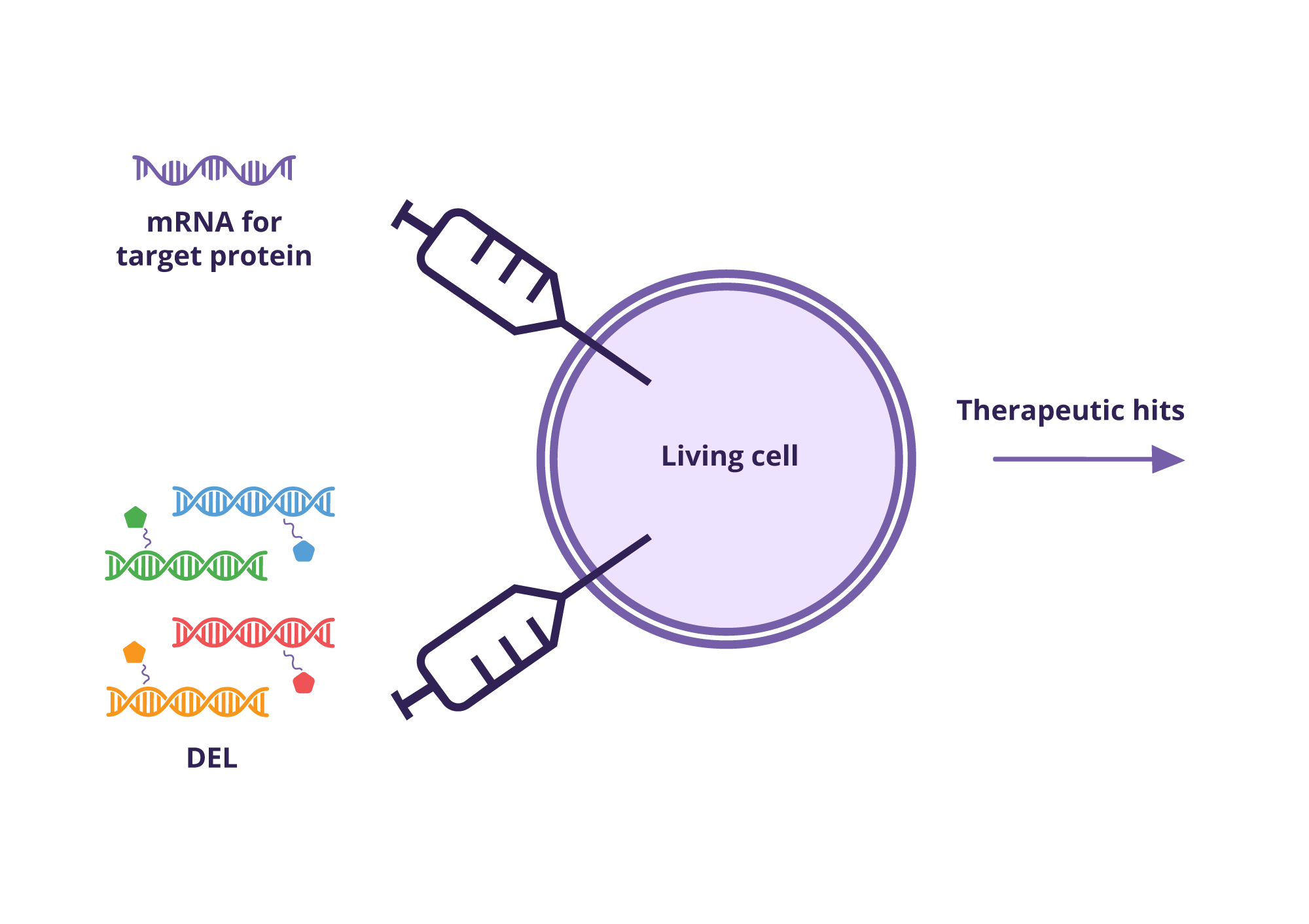
Test compounds or compound libraries are introduced into the cellular system. In advanced screening approaches, such as DNA-encoded library (DEL) screening, compounds may be delivered together with DNA constructs that enable downstream identification.

3. Incubation and interaction

Cells are incubated under controlled conditions to allow compounds to interact with the target protein in its native cellular context. This step enables binding events that depend on proper protein folding, membrane insertion, and intracellular environment.

4. Binding stabilization and signal generation

Depending on the assay design, binding events are detected through functional readouts (e.g., fluorescence or luminescence) or through molecular enrichment strategies. In binding-based systems, conditions are applied to selectively retain compounds that interact with the target.

5. Cell lysis and sample processing

Cells are lysed to recover relevant biological material. In some workflows, additional processing steps—such as DNA ligation, amplification, or labeling—are performed to prepare samples for analysis.

6. Data collection and analysis

Signals or molecular identifiers are collected and analyzed to identify active compounds. In DEL-based approaches, next-generation sequencing is used to decode DNA barcodes associated with enriched compounds. (Gironda-Martínez et al., 2021).

7. Hit validation and follow-up

Identified hits are validated through orthogonal assays to confirm binding and assess functional activity. This step is essential to ensure biological relevance and reproducibility.

Innovative Applications and Case Studies in Cell-Based Assays

Transformative Applications in Drug Discovery and Toxicology

Modern cell-based assays have become essential tools in drug discovery by enabling the assessment of compound–target interactions in a biologically relevant context. Unlike traditional in vitro systems that rely on purified proteins or artificial membranes, cell-based assays preserve native features such as protein folding, membrane insertion, and subcellular compartmentalization. This makes them particularly valuable for screening difficult target classes like ion channels, GPCRs, and intracellular protein–protein interactions.

An important innovation in this space is the use of binding-based cell assays, which focus on compound engagement rather than downstream functional effects. For example, small molecules that act as molecular glues — stabilizing multiprotein complexes — require intact structural interfaces that are often only preserved in cellular environments.  (Moffat et al) Such assays are increasingly used to validate hits from DNA-encoded library (DEL) screens and fragment libraries, helping to triage candidates based on selective and physiologically meaningful interactions.

In early toxicology, cell-based assays offer predictive insight into safety profiles. By measuring compound interactions in disease-relevant membrane environments or transport-sensitive contexts, these systems complement traditional ADMET profiling with mechanistic data on off-target binding or membrane disruption (Xu & Li, 2007).

Real-World Case Studies

  • 3D cancer spheroid assays have demonstrated the ability to detect subtle cytostatic effects and morphological alterations not observable in traditional monolayer cultures, thus improving relevance for oncology models [Sirenko et al., 2015].
  • High-content screening (HCS) platforms have enabled the discovery of pathway-selective modulators in complex systems such as neurodegenerative disease models, where multiparametric image analysis reveals compound-specific phenotypes beyond cell viability.

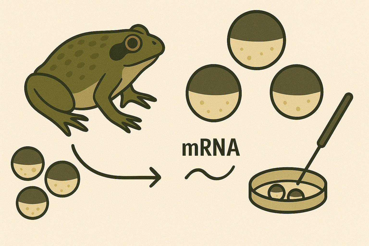
Oocyte-based binding assays, including Vipergen’s cellular Binder Trap Enrichment (cBTE) https://www.vipergen.com/cellular-binder-trap-enrichment/ screening, allow DNA-encoded libraries to be screened directly in live Xenopus laevis oocytes, a well-established system for heterologous protein expression and electrophysiological measurements (Stühmer, 1992). These cells express proteins in a native-like environment, enabling detection of binding events for structurally complex targets that are often difficult to purify or assay using classical methods.

Traditional vs. Modern Assay Techniques

While traditional assays often relied on isolated enzymes or purified receptors in artificial systems, modern cell-based platforms increasingly assess molecular interactions in a biologically relevant context. The use of intact, live cells — including oocyte-based systems — allows researchers to capture aspects of protein folding, subcellular localization, and conformational dynamics that are essential for meaningful interaction profiling.

This is particularly important in the study of challenging targets such as protein–protein interactions and molecular glues, where binding interfaces may only be preserved in native-like environments. Screening directly in living cells enables the identification of small molecules that stabilize or disrupt multiprotein complexes — an area of growing importance in next-generation drug discovery.

Emerging Trends and Innovations

Recent innovations in cell-based screening include:

  • Miniaturized organoid systems for modelling tissue-specific drug responses in oncology and regenerative medicine
  • Machine learning–enhanced high-content imaging, which enables phenotypic clustering and multiparametric analysis of cellular responses (Caicedo et al., 2017)
  • Label-free detection technologies, such as impedance-based readouts and thermal shift assays
  • Cell-based binding assays in intact membrane environments, including oocyte-based screening platforms that preserve lipid bilayers and protein folding — enabling detection of ligand interactions with GPCRs, ion channels, and protein–protein interfaces
  • Assays targeting molecular glues and induced proximity, reflecting the growing interest in stabilizers of protein–protein interactions and targeted degradation mechanisms (Schapira et al., 2019)

These innovations illustrate a broader shift: from purely functional or biochemical readouts to mechanism-informed screening in biologically relevant systems.

Industry Perspectives

Robust and reproducible cell-based assays are essential for de-risking early-stage compounds. The convergence of structural biology, binding data, and cellular context has transformed how lead molecules are selected and optimized. As the field moves toward targeting previously “undruggable” proteins — such as intracellular scaffolds or dynamic complexes — the role of physiologically relevant, mechanism-specific assays becomes increasingly central to pipeline success.

For projects that must move quickly from simple binding detection to functional assays, these technologies supply chemically verified starting points already selected in a relevant cellular or solution-based context. https://www.vipergen.com/services/

Conclusion

Cell-based assays have firmly established themselves as indispensable tools in modern scientific research and drug discovery by enabling the assessment of molecular interactions in biologically relevant systems

Advances in assay formats — from 3D cultures and high-content imaging to oocyte-based binding platforms — have significantly enhanced the physiological fidelity and predictive power of these systems. This makes them especially valuable for examining challenging targets like GPCRs, ion channels, and protein–protein interactions.

As therapeutic discovery increasingly focuses on difficult and dynamic targets, the role of robust, mechanism-informed, and physiologically relevant screening platforms will only grow.

Vipergen’s contribution to cell-based screening

Cellular Binder Trap Enrichment (cBTE) performs the DEL screen inside living cells, so binders are identified under physiologically relevant conditions without the need for purified protein. the workflow relies on the so-called YoctoReactor libraries https://www.vipergen.com/yoctoreactor-yr-libraries/—hundreds of millions of well-defined, DNA-tagged compounds synthesised in a single tube—reducing false positives and making hit resynthesis manageable.  

For projects that must move quickly from simple binding detection to functional assays, these technologies supply chemically verified starting points already selected in a relevant cellular or solution-based context. https://www.vipergen.com/services/

Frequently Asked Questions

  • What is the difference between cell-based and biochemical assays?

    Cell-based assays measure how compounds and / or biological processes behave in living cells, preserving a more realistic biological context such as protein interactions, membrane environment, and cellular signaling pathways. In contrast, biochemical assays are performed on isolated molecules or proteins in vitro. Offering a mere controlled setup but less biologically relevant data.

  • What are the main types of cell-based assays?

    Common types of cell-based assays include reporter gene assays, viability and cytotoxicity assays, proliferation assays, apoptosis assays, and functional assays such as calcium flux or electrophysiology. High-content imaging assays are also widely used when more detailed information about cellular behaviour or phenotype is needed.

  • How long does a cell-based assay take?

    The timeline depends on assay type and biological system, but typically ranges from a few hours to several days. Assays requiring cell growth, differentiation, or longer incubation periods usually take longer than standard biochemical assays.

  • What equipment is needed for cell-based assays?

    Most Cell-based assays typically require cell culture facilities, such as incubators, biosafety cabinets, and appropriate culture media. Detection systems vary depending on the assay and may include plate readers for fluorescence or luminescence, microscopes for imaging, or specialized electrophysiology equipment.

  • Why are cell-based assays important in drug discovery?

    Cell-based assays play an important role in drug discovery because they show how a compound behaves in a biologically relevant environment. They provide insights into cellular responses, target engagement, and potential toxicity earlier in the process. As a result, they often provide a better indication of whether observed effects translate to in vivo systems.

References

  1. Swinney, D. C., & Anthony, J. (2011). How were new medicines discovered? Nat Rev Drug Discov, 10(7), 507–519. https://doi.org/10.1038/nrd3480
  2. Sirenko, O., Mitlo, T., Hesley, J., Luke, S., Owens, W., & Cromwell, E. F. (2015). High-content assays for characterizing the viability and morphology of 3D cancer spheroid cultures. Assay Drug Dev Technol, 13(7), 402–414. https://doi.org/10.1089/adt.2015.655
  3. Moffat, J. G., Vincent, F., Lee, J. A., Eder, J., & Prunotto, M. (2017). Opportunities and challenges in phenotypic drug discovery: An industry perspective. Nat Rev Drug Discov, 16(8), 531–543. https://doi.org/10.1038/nrd.2017.111
  4. Dunlop, J., Bowlby, M., Peri, R., Vasilyev, D., & Arias, R. (2008). High-throughput electrophysiology: An emerging paradigm for ion-channel screening and physiology. Nat Rev Drug Discov, 7(4), 358–368. https://doi.org/10.1038/nrd2552
  5. Stühmer, W. (1992). Electrophysiological recording from Xenopus oocytes. Methods in Enzymology, 207, 319–339. https://doi.org/10.1016/0076-6879(92)07026-
  6. Zhang, J.H., Chung, T.D., & Oldenburg, K.R. (1999). A Simple Statistical Parameter for Use in Evaluation and Validation of High Throughput Screening Assays. J Biomol Screen, 4(2), 67–73. https://doi.org/10.1177/108705719900400206
  7. Xu, J. J., Henstock, P. V., Dunn, M. C., Smith, A. R., Chabot, J. R., & de Graaf, D. (2008). Cellular imaging predictions of clinical drug-induced liver injury. Toxicological Sciences, 105(1), 97–105.
    DOI: https://doi.org/10.1093/toxsci/kfn109
  8. Caicedo, J.C., et al. (2017). Data-analysis strategies for image-based cell profiling. Nature Methods, 14, 849–863. https://doi.org/10.1038/nmeth.4397
  9. Schapira, M., et al. (2019). Targeted protein degradation: expanding the toolbox. Nature Reviews Drug Discovery, 18(12), 949–963. https://doi.org/10.1038/s41573-019-0047-y
  1. Wei, F., Wang, S., & Gou, X. (2021). A review for cell-based screening methods in drug discovery. Biophysics Reports, 7(6), 504–516. https://doi.org/10.52601/bpr.2021.210042
  2. Gironda-Martínez, A., Donckele, E. J., Samain, F., Neri, D., & DNA-Encoded Chemical Libraries Network. (2021). DNA-Encoded Chemical Libraries: A Comprehensive Review with Successful Stories and Future Challenges. ACS Pharmacology & Translational Science, 4(4), 1265–1279. https://doi.org/10.1021/acsptsci.1c00118

Do you have an inquiry?

Contact us today and explore partnership opportunities.

Get In Touch

Related Services

Service
Small molecule drug discovery for even hard-to-drug targets – identify inhibitors, binders and modulators
Molecular Glue Direct
PPI Inhibitor Direct
Integral membrane proteins
Specificity Direct – multiplexed screening of target and anti-targets
Express – optimized for fast turn – around-time
Snap – easy, fast, and affordable

Continue reading

Molecular Display Systems in Drug Discovery: Principles, Platforms, and Applications

Phage Display Technology: Principles, Libraries, and Applications in Drug Discovery

Get In Touch

Introduction

Phage display technology is a molecular screening method in which bacteriophages are engineered to display peptides or proteins on their surfaces while carrying the encoding DNA internally. This direct genotype–phenotype linkage enables the construction of phage display libraries with up to 10¹⁰ variants, allowing high-throughput screening for molecules with desirable binding properties. The concept was pioneered in 1985 by George P. Smith, who demonstrated that foreign peptides could be fused to phage coat proteins and displayed in a heritable fashion.

Since its introduction, phage display has become a cornerstone of molecular discovery, particularly in the fields of antibody engineering, peptide ligand discovery, epitope mapping, and vaccine development. The technology’s ability to generate human therapeutic antibodies has had profound impact, with adalimumab (Humira) standing as a landmark example of a phage display–derived monoclonal antibody approved for clinical use [1,2].

Despite the emergence of other display and library technologies such as mRNA display [3,4] and DNA-encoded libraries (DELs) [5-8], phage display remains highly relevant due to its robustness, scalability, and direct compatibility with therapeutic antibody pipelines. In this review, we examine the principles, methods, and applications of phage display technology, with extended comparisons to mRNA display and DEL platforms.

How Phage Display Works

Genotype–phenotype linkage

The foundation of phage display technology is the physical coupling of genotype (encoding DNA) with phenotype (displayed peptide or protein). A foreign DNA sequence is inserted into the gene encoding a phage coat protein, resulting in the fusion of the encoded peptide/protein to the coat protein displayed on the viral surface. The corresponding DNA resides inside the phage particle, ensuring that each displayed molecule is linked to its genetic blueprint.

Common phage systems

Several bacteriophage systems are employed in phage display:

  • M13 filamentous phage: The most widely used system. The minor coat protein pIII (3–5 copies) is suited for displaying larger proteins such as antibody fragments, while the major coat protein pVIII (~2700–3000 copies) supports high-valency display of short peptides.
  • T7 phage: Offers greater robustness and can display larger proteins without requiring secretion through the bacterial membrane.
  • T4 phage: Capable of displaying very large proteins and multivalent constructs.
  • λ phage: Less common but useful for certain protein formats.

Helper phages and phagemid vectors are often employed in M13 systems. Phagemids carry the display construct, while helper phages provide the structural proteins needed for phage assembly.

Biopanning cycle

The process of phage display screening, also known as biopanning, involves iterative rounds of affinity selection:

  1. Library incubation – A large phage display library is exposed to an immobilised target (purified protein, peptide, or even whole cells).
  2. Washing – Non-binding phages are washed away under increasingly stringent conditions.
  3. Elution – Bound phages are eluted, often by pH shift, enzymatic cleavage, or competitive ligands.
  4. Amplification – Eluted phages are amplified in E. coli, regenerating the pool for the next round.
  5. Enrichment – After 3–5 rounds, high-affinity binders are enriched and sequenced.

Figure 1: Biopanning cycle

What Is a Phage Display Library?

A phage display library is a diverse collection of bacteriophages, each presenting a unique peptide, antibody fragment, or protein variant on its surface. The diversity of a library can reach 10⁹–10¹⁰ unique members, significantly enhancing the probability of identifying rare, high-affinity binders.

Types of phage display libraries

  • Peptide libraries:
    • Synthetic libraries use randomised oligonucleotides to generate peptides with controlled diversity.
    • Natural libraries use DNA fragments from biological sources.
    • Semi-synthetic libraries combine both strategies.
  • Antibody libraries:
    • Naïve libraries are derived from B-cell repertoires of healthy donors, constructed via splice-by-overlap extension PCR, providing broad diversity.
    • Immune libraries originate from immunised donors and typically yield higher-affinity clones against the specific antigen.
    • Synthetic antibody libraries incorporate designed CDR (complementarity-determining region) sequences.
  • Fragment formats:
    • scFv (single-chain variable fragments)
    • Fab (fragment antigen-binding)
    • VH and nanobody formats

Vector systems

Applications of Phage Display

Phage display technology has broad applications across biomedical research and drug discovery:

Antibody engineering

Phage display revolutionised antibody discovery by enabling the generation of fully human antibodies without the need for hybridoma technology. Adalimumab (Humira) is a landmark product developed using phage display [1,2]. Numerous therapeutic antibodies across oncology, inflammation, and infectious diseases have since been identified using phage display antibody libraries.

Peptide ligand discovery

Phage display peptide libraries are valuable tools for identifying binding motifs against receptors, enzymes, and protein–protein interaction interfaces. Short peptide ligands discovered via phage display have been used as leads for therapeutics, diagnostics, and targeting agents.

Epitope mapping and vaccine design

Epitope mapping with phage display identifies the precise binding sites of antibodies on antigens, aiding rational vaccine design and immunodiagnostic assay development.

Emerging applications

  • Nanobody generation: Selection of VH domains and nanobody scaffolds against membrane proteins.
  • Cell-surface selection: Direct screening against intact cells for immuno-oncology targets.
  • Diagnostic peptides: For example, HER3P1 peptide has been developed for imaging HER3 expression in tumors.

Figure 2: Application of phage display

Advantages and Limitations of Phage Display

Advantages

  • Extremely large library sizes (up to ~10¹⁰ variants) 
  • Direct genotype–phenotype linkage
  • Straightforward amplification in bacteria
  • Cost-effective and scalable
  • Rapid identification of high-affinity ligands

Limitations

  • Restricted to peptides and proteins (no small-molecule diversity)
  • Post-translationally modified proteins are difficult to display
  • Large proteins may not fold correctly in the phage context
  • cDNA libraries may contain stop codons or non-functional clones

Comparison with Other Display Platforms

mRNA Display

mRNA display is a cell-free in vitro translation system where peptides are covalently linked to their encoding mRNA via puromycin [3]. Key features include:

  • Library size: Up to 10¹³ members, surpassing phage libraries.
  • Chemical diversity: Incorporation of some non-natural amino acids and macrocyclic scaffolds [4].
  • Applications: Particularly suited for targeting protein–protein interaction (PPI) interfaces.

DNA-Encoded Library (DEL) Technology

DEL combines combinatorial chemistry with DNA barcoding [5-7]. Features include:

  • Library size: 10⁶–10¹² small molecules.
  • Chemical diversity: Very high; compatible with drug-like small molecules.
  • Screening: Pooled libraries incubated with targets, followed by PCR enrichment and NGS readout [6].
  • Applications: Small-molecule discovery, especially for enzymatic targets, PPIs, and molecular glues.
  • Integration with AI/ML: Machine learning enhances predictive power and hit triaging [8].
Parameter Phage Display mRNA Display DEL Technology
Molecular class Peptides, proteins Peptides, macrocycles Small molecules and peptides
Library size 109–1011 1012–1013 106–1012
Chemical diversity Limited Moderate to high Very high
In vitro compatibility Partial Full Full
Target types Proteins, cells Proteins, PPIs Proteins, enzymatic targets, molecular glues, PPIs

Table 1: Comparative table of phage display, mRNA display, and DEL

Trends in Commercial Use and Collaboration

Pharmaceutical companies increasingly rely on collaborations with contract research organizations (CROs) and platform providers to access proprietary phage display libraries, custom library construction, and data analysis expertise. Outsourcing offers:

  • Access to validated and proprietary antibody/peptide libraries
  • Customisation for specific targets (including membrane proteins and GPCRs)
  • Data interpretation, clustering, and selectivity profiling

Integration with structure-based drug design, cryo-EM, and computational modelling further accelerates lead optimization. AI-augmented approaches, particularly in DEL screening, are increasingly being applied to phage display datasets as well.

Vipergen’s DNA-Encoded Library Screening Platform

Although this review is centred on phage display, complementary technologies such as DNA-encoded library screening services play a vital role in small-molecule discovery. At Vipergen, we provide custom DEL screening solutions with:

  • Modular library design with privileged scaffolds and novel chemotypes
  • Screening against challenging targets, including membrane proteins and in intact cells
  • Integrated SAR analysis and cheminformatics clustering
  • Multiplexed selectivity profiling against target and anti-targets

For researchers pursuing oncology, GPCR ligands, or chemical probes for validation, our DEL screening services provide a high-throughput, scalable platform that complements phage display–based biologics discovery.

See services

Conclusion

Phage display technology remains one of the most influential methods in molecular discovery. By harnessing genotype–phenotype linkage, phage display libraries enable rapid screening of billions of peptides, proteins, and antibody fragments. The method’s impact on antibody therapeutics, peptide discovery, and epitope mapping underscores its continuing relevance, even as complementary technologies such as mRNA display and DEL broaden the molecular space accessible to researchers.

As library construction methods, biopanning strategies, and computational tools evolve, phage display screening will continue to serve as a cornerstone of biologics discovery while integrating seamlessly with next-generation discovery platforms. Its combination of scalability, cost effectiveness, and clinical track record ensures that phage display remains indispensable in the next era of precision drug discovery.

References

  1. McCafferty J, Griffiths AD, Winter G, Chiswell DJ. Phage antibodies: filamentous phage displaying antibody variable domains. Nature. 1990;348(6301):552–554. https://doi.org/10.1038/348552a0
  2. Winter G, Griffiths AD, Hawkins RE, Hoogenboom HR. Making antibodies by phage display technology. Annu Rev Immunol. 1994;12:433–455. https://doi.org/10.1146/annurev.iy.12.040194.002245
  3. Roberts RW, Szostak JW. RNA–peptide fusions for the in vitro selection of peptides and proteins. Proc Natl Acad Sci USA. 1997;94(23):12297–12302. https://doi.org/10.1073/pnas.94.23.12297
  4. Huang Y, Wiedmann MM, Suga H. RNA Display Methods for the Discovery of Bioactive Macrocycles. Chem Rev. 2019;119(17):10360–10391. https://doi.org/10.1021/acs.chemrev.8b00430
  5. Peterson AA, Liu DR. Small-molecule discovery through DNA-encoded libraries. Nat Rev Drug Discov. 2023;22(9):699–722. https://doi.org/10.1038/s41573-023-00713-6
  6. Favalli N, Bassi G, Scheuermann J, Neri D. DNA-encoded chemical libraries – achievements and remaining challenges. FEBS Lett. 2018;592(17):2168-2180. https://doi.org/10.1002/1873-3468.13068
  7. Mason JW, Wang Y, et al. DNA-encoded library-enabled discovery of proximity-inducing small molecules. Nat Chem Biol. 2024;20:170–179. https://doi.org/10.1038/s41589-023-01458-4
  8. McCloskey K, Sigel EA, et al. Machine learning on DNA-encoded libraries: A new paradigm for hit-finding. J Med Chem. 2020;63(16):8857-8866. https://doi.org/10.1021/acs.jmedchem.0c00452

Do you have an inquiry?

Contact us today and explore partnership opportunities.

Get In Touch

Related Services

Service
Small molecule drug discovery for even hard-to-drug targets – identify inhibitors, binders and modulators
Molecular Glue Direct
PPI Inhibitor Direct
Integral membrane proteins
Specificity Direct – multiplexed screening of target and anti-targets
Express – optimized for fast turn – around-time
Snap – easy, fast, and affordable

Continue reading

Xenopus Oocytes in Human Protein Research and Drug Discovery

Xenopus Oocytes in Human Protein Research and Drug Discovery

Get In Touch

Historical Background and Core Advantages

The utilization of frog oocytes, particularly those from the African clawed frog (Xenopus laevis), has become important in the study of human proteins. These oocytes, or immature egg cells, provide a robust and versatile system for the expression and functional analysis of various proteins, including ion channels, transporters, and receptors (Ivorra et al., 2022). Due to their unique biological properties and ease of manipulation, Xenopus oocytes have become an invaluable tool in biomedical research.
The scientific use of Xenopus oocytes began in the mid-20th century and was revolutionized in the 1970s with the development of mRNA microinjection techniques. Since then, these cells have been employed as a key system for in vitro expression and analysis of proteins from diverse species.

Why Xenopus oocytes?

– Natural protein synthesis machinery: Supports complex folding and post-translational modifications similar to human cells
– Wide expression capabilities: Can express challenging human proteins that do not fold or function properly in other systems
– Year-round availability: Commercial suppliers provide high-quality oocytes consistently
– Large size (1.0–1.3 mm): Easy to handle and manipulate

These features make them highly effective for studying human membrane proteins, intracellular signaling pathways, and receptor-ligand interactions.

Functional Expression and Assays

After mRNA injection, functional assays allow detailed investigation of expressed proteins. Some key methods include:
– Patch-clamp electrophysiology: Measures ion channel activity with high precision
– Radiolabeled uptake assays: Quantifies transporter function
– Ligand binding and pharmacology assays: Evaluates receptor interactions with small molecules
– DEL screening: Screening of DNA Encoded Libraries (DELs) in living cells

Applications in Human Protein and Disease Research

Xenopus oocytes are widely used to model human diseases and understand the function of human proteins, especially in the nervous system, cardiovascular system, and metabolism.

Neurological Targets

Xenopus oocytes have played a central role in characterizing key targets involved in central nervous system (CNS) disorders. Functional expression of human serotonin (5-HT) receptors, GABA_A and glutamate receptors, as well as voltage-gated potassium (Kv) and sodium (Na_v) channels, has allowed researchers to elucidate biophysical properties and pharmacological profiles critical to neuropsychiatric and neurodegenerative drug discovery. This model enables precise dose-response assessments, agonist/antagonist screening, and evaluation of modulatory compounds under near-physiological conditions. For example, selective serotonin receptor modulators, developed for mood and anxiety disorders, have been validated in oocyte assays due to their ability to maintain native receptor conformations and G-protein coupling properties (O’Connor et al., 2023).

Cystic Fibrosis and Ion Channelopathies

The functional analysis of mutated CFTR (cystic fibrosis transmembrane conductance regulator) variants in Xenopus oocytes has significantly advanced the understanding of genotype-phenotype correlations in cystic fibrosis. The system allows for rapid assessment of CFTR activity via chloride conductance assays, providing a robust platform to evaluate therapeutic candidates, including small-molecule potentiators and correctors. Oocytes expressing mutant CFTR can be treated with test compounds to validate functional rescue, a step critical in preclinical screening workflows (Kvist et al., 2011).

Diabetes and Metabolic Disorders

By co-expressing human insulin receptors (IR) with glucose transporters (such as GLUT4), Xenopus oocytes have provided an experimental model to reconstruct insulin signaling cascades. This approach has been used to study insulin-stimulated hexose uptake and to validate the effect of insulin-mimetic compounds and insulin pathway modulators. Such assays have been used to evaluate the pharmacological activity of candidate compounds targeting Type 2 diabetes and insulin resistance (Vera & Rosen, 1990).

Cardiovascular Research

Xenopus oocytes have been used to study the electrophysiology and pharmacology of cardiac ion channels such as hERG (human Ether-à-go-go-Related Gene) potassium channels, which are critical for cardiac repolarization. Functional expression of hERG in oocytes supports safety pharmacology screening by identifying compounds that may cause QT prolongation, a known risk factor for drug-induced arrhythmias. (Dascal, 1987 Yang et al., 2025).

Oncology and Cell Signaling

The oocyte model enables detailed dissection of mitogenic and apoptotic pathways by expressing components of the MAPK, PI3K/AKT, and other signal transduction pathways. Researchers have used Xenopus oocytes to analyze kinase activity, post-translational modifications, and drug-induced pathway modulation. This model supports the functional validation of small molecule inhibitors targeting oncogenic kinases and phosphatases and aids in the identification of lead compounds for cancer therapy (Dupré et al., 2011).

DNA Encoded Library (DEL) Screening in Living Cells

An innovative leap in drug discovery is the use of Xenopus oocytes for DEL screening in live cells, pioneered by Vipergen in 2021. This approach allows for high-throughput screening against proteins in their native cellular context (Petersen et al., 2021). 

Cellular Binder Trap Enrichment

This technology is amenable for several target classes and offered by Vipergen as a DEL screening service

See services

Conclusion

Xenopus laevis oocytes are a cornerstone of human protein research and modern drug discovery. Their utility spans basic biology, disease modeling, and compound screening including in-cell DEL screening.

References

De Robertis, E. M., & Gurdon, J. B. (2021). A Brief History of Xenopus in Biology. Cold Spring Harbor Protocols, https://doi.org/10.1101/pdb.top107615

Dascal, N. (1987). The Use of Xenopus Oocytes for the Study of Ion Channels. Critical Reviews in Biochemistry, 22(4), 317. https://doi.org/10.3109/10409238709086960

Dupré, A., Haccard, O., & Jessus, C. (2011). Mos in the Oocyte: How to Use MAPK Independently of Growth Factors and Transcription to Control Meiotic Divisions. Journal of Signal Transduction 1, Article ID 350412 https://doi.org/10.1155/2011/350412

Gurdon, J.B., Lane, C.D., et al. (1971). Use of frog eggs and oocytes for the study of messenger RNA and its translation in living cells. Nature 233, 177. https://doi.org/10.1038/233177a0

Ivorra, I, Alberola-Die, A., et al. (2022). Xenopus Oocytes as a Powerful Cellular Model to Study Foreign Fully-Processed Membrane Proteins. Membranes 12, 986. https://doi.org/10.3390/membranes12100986

Kvist, T., Hansen, K. B., & Bräuner-Osborne, H. (2011). The use of Xenopus oocytes in drug screening. Expert Opinion on Drug Discovery, 6(2), 141. https://doi.org/10.1517/17460441.2011.546396

Nutt, L. (2012). The Xenopus oocyte: A model for studying the metabolic regulation of cancer cell death. Seminars in Cell and Developmental Biology 23, 412. https://doi.org/10.1016/j.semcdb.2012.03.015

O’Connor, E. C., Kambara, K., & Bertrand, D. (2023). Advancements in the use of Xenopus oocytes for modelling neurological disease for novel drug discovery. Expert Opinion on Drug Discovery, 19(2), 173. https://doi.org/10.1080/17460441.2023.2270902

Pehl, U., Leisgen, C., Gampe, K., & Guenther, E. (2004). Automated higher-throughput compound screening on ion channel targets based on the Xenopus laevis oocyte expression system. Assay and Drug Development Technologies, 2(5), 515. https://doi.org/10.1089/adt.2004.2.515

Petersen, L. K. et. Al., 2021, Screening of DNA-Encoded Small Molecule Libraries inside a Living Cell, J. Am. Chem. Soc., 143, 7, 2751. https://doi.org/10.1021/jacs.0c09213

Vera, J. C., & Rosen, O. M. (1990). Reconstitution of an insulin signaling pathway in Xenopus laevis oocytes: Coexpression of a mammalian insulin receptor and three different mammalian hexose transporters. Molecular and Cellular Biology, 10(2), 743. https://doi.org/10.1128/mcb.10.2.743-751

Do you have an inquiry?

Contact us today and explore partnership opportunities.

Get In Touch

Related Services

Service
Small molecule drug discovery for even hard-to-drug targets – identify inhibitors, binders and modulators
Molecular Glue Direct
PPI Inhibitor Direct
Integral membrane proteins
Specificity Direct – multiplexed screening of target and anti-targets
Express – optimized for fast turn – around-time
Snap – easy, fast, and affordable

Continue reading